 Movie
Movie Controller

[English] 日本語
 Yorodumi
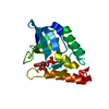
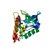
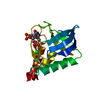
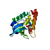
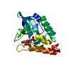
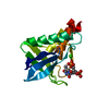
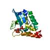
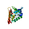
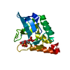
- PDB-6xsc: Crystal structure of Staphylococcal nuclease variant Delta+PHS V2... -
+ Open data

- Basic information
| Entry | Database: PDB / ID: 6xsc | ||||||
|---|---|---|---|---|---|---|---|
| Title | Crystal structure of Staphylococcal nuclease variant Delta+PHS V23T at cryogenic temperature | ||||||
 Components Components | Thermonuclease | ||||||
 Keywords Keywords | HYDROLASE / nuclease / hyperstable / OB-fold / internal polar group / pdTp | ||||||
| Function / homology |  Function and homology information Function and homology informationmicrococcal nuclease / 3' overhang single-stranded DNA endonuclease activity / nucleic acid binding / extracellular region / membrane / metal ion binding Similarity search - Function | ||||||
| Biological species |   Staphylococcus aureus (bacteria) Staphylococcus aureus (bacteria) | ||||||
| Method |  X-RAY DIFFRACTION / MOLECULAR REPLACEMENT / Resolution: 2 Å X-RAY DIFFRACTION / MOLECULAR REPLACEMENT / Resolution: 2 Å | ||||||
 Authors Authors | Robinson, A.C. / Schlessman, J.L. / Garcia-Moreno E., B. | ||||||
| Funding support |  United States, 1items United States, 1items
| ||||||
 Citation Citation | Journal: To be Published Title: Crystal structure of Staphylococcal nuclease variant Delta+PHS V23T at cryogenic temperature Authors: Robinson, A.C. / Schlessman, J.L. / Garcia-Moreno E., B. | ||||||
| History |
|
- Structure visualization
| Structure viewer | Molecule: MolmilJmol/JSmol |
|---|
- Downloads & links
-Download
| PDBx/mmCIF format | 6xsc.cif.gz | 44 KB | Display | PDBx/mmCIF format |
|---|---|---|---|---|
| PDB format | pdb6xsc.ent.gz | 27.2 KB | Display | PDB format |
| PDBx/mmJSON format | 6xsc.json.gz | Tree view | PDBx/mmJSON format | |
| Others |  Other downloads Other downloads |
-Validation report
| Arichive directory | https://data.pdbj.org/pub/pdb/validation_reports/xs/6xscftp://data.pdbj.org/pub/pdb/validation_reports/xs/6xsc | HTTPS FTP |
|---|
-Related structure data
| Related structure data | 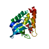 3bdcS S: Starting model for refinement |
|---|---|
| Similar structure data |


-Links
 PDBj
PDBj- Assembly
| Deposited unit | 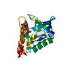
| ||||||||
|---|---|---|---|---|---|---|---|---|---|
| 1 |
| ||||||||
| Unit cell |
|
-Components
| #1: Protein | Mass: 16145.436 Da / Num. of mol.: 1 / Fragment: UNP residues 83-231 / Mutation: V23T/G50F/V51N/P117G/H124L/S128A/Del44-49 Source method: isolated from a genetically manipulated source Source: (gene. exp.) Staphylococcus aureus (bacteria) / Gene: nuc / Plasmid: pET24a+ / Production host: |
|---|---|
| #2: Chemical | ChemComp-THP /   Type: DNA linking / Mass: 402.188 Da / Num. of mol.: 1 / Source method: obtained synthetically / Formula: C10H16N2O11P2 Type: DNA linking / Mass: 402.188 Da / Num. of mol.: 1 / Source method: obtained synthetically / Formula: C10H16N2O11P2 |
| #3: Chemical | ChemComp-CA /   Mass: 40.078 Da / Num. of mol.: 1 / Source method: obtained synthetically / Formula: Ca Mass: 40.078 Da / Num. of mol.: 1 / Source method: obtained synthetically / Formula: Ca |
| #4: Water | ChemComp-HOH /  Mass: 18.015 Da / Num. of mol.: 43 / Source method: isolated from a natural source / Formula: H2O Mass: 18.015 Da / Num. of mol.: 43 / Source method: isolated from a natural source / Formula: H2O |
-Experimental details
-Experiment
| Experiment | Method: X-RAY DIFFRACTION / Number of used crystals: 1 |
|---|
- Sample preparation
| Crystal | Density Matthews: 2.21 Å3/Da / Density % sol: 44.41 % |
|---|---|
| Crystal grow | Temperature: 277 K / Method: vapor diffusion, hanging drop / pH: 8 Details: 20% MPD, 25 mM potassium phosphate, calcium chloride |
-Data collection
| Diffraction | Mean temperature: 110 K / Serial crystal experiment: N |
|---|---|
| Diffraction source | Source: SEALED TUBE / Type: Agilent SuperNova / Wavelength: 1.54 Å |
| Detector | Type: AGILENT ATLAS CCD / Detector: CCD / Date: Mar 16, 2018 |
| Radiation | Protocol: SINGLE WAVELENGTH / Monochromatic (M) / Laue (L): M / Scattering type: x-ray |
| Radiation wavelength | Wavelength: 1.54 Å / Relative weight: 1 |
| Reflection | Resolution: 2→38.04 Å / Num. obs: 9461 / % possible obs: 99.9 % / Observed criterion σ(F): 0 / Observed criterion σ(I): 0 / Redundancy: 10.1 % / Biso Wilson estimate: 35.3 Å2 / Rmerge(I) obs: 0.064 / Net I/σ(I): 37.26 |
| Reflection shell | Resolution: 2→2.07 Å / Redundancy: 6.1 % / Rmerge(I) obs: 0.276 / Mean I/σ(I) obs: 4.79 / Num. unique obs: 961 / % possible all: 100 |
- Processing
| Software |
| ||||||||||||||||||||||||||||||||||||||||||||||||||||||||||||
|---|---|---|---|---|---|---|---|---|---|---|---|---|---|---|---|---|---|---|---|---|---|---|---|---|---|---|---|---|---|---|---|---|---|---|---|---|---|---|---|---|---|---|---|---|---|---|---|---|---|---|---|---|---|---|---|---|---|---|---|---|---|
| Refinement | Method to determine structure: MOLECULAR REPLACEMENT Starting model: PDB entry 3BDC Resolution: 2→38.04 Å / Cor.coef. Fo:Fc: 0.951 / Cor.coef. Fo:Fc free: 0.934 / SU B: 6.433 / SU ML: 0.165 / SU R Cruickshank DPI: 0.2077 / Cross valid method: THROUGHOUT / σ(F): 0 / ESU R: 0.208 / ESU R Free: 0.183 Details: HYDROGENS HAVE BEEN ADDED IN THE RIDING POSITIONS U VALUES : REFINED INDIVIDUALLY
| ||||||||||||||||||||||||||||||||||||||||||||||||||||||||||||
| Solvent computation | Ion probe radii: 0.8 Å / Shrinkage radii: 0.8 Å / VDW probe radii: 1.2 Å | ||||||||||||||||||||||||||||||||||||||||||||||||||||||||||||
| Displacement parameters | Biso max: 65.59 Å2 / Biso mean: 32.725 Å2 / Biso min: 17.86 Å2
| ||||||||||||||||||||||||||||||||||||||||||||||||||||||||||||
| Refinement step | Cycle: final / Resolution: 2→38.04 Å
| ||||||||||||||||||||||||||||||||||||||||||||||||||||||||||||
| Refine LS restraints |
| ||||||||||||||||||||||||||||||||||||||||||||||||||||||||||||
| LS refinement shell | Resolution: 2→2.052 Å / Rfactor Rfree error: 0 / Total num. of bins used: 20
|
