 Movie
Movie Controller


+ Open data

- Basic information
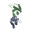
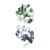
| Entry | Database: PDB / ID: 6spv | ||||||
|---|---|---|---|---|---|---|---|
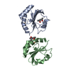
| Title | Crystal structure of PDZ1-2 from PSD-95 | ||||||
 Components Components | Disks large homolog 4 | ||||||
 Keywords Keywords | STRUCTURAL PROTEIN / PSD-95 fragment / clustering / protein-complex / receptor-binding / membrane associated | ||||||
| Function / homology |  Function and homology information Function and homology informationLGI-ADAM interactions / P2Y1 nucleotide receptor binding / beta-1 adrenergic receptor binding / neuroligin family protein binding / regulation of grooming behavior / NrCAM interactions / synaptic vesicle maturation / positive regulation of neuron projection arborization / receptor localization to synapse / establishment or maintenance of epithelial cell apical/basal polarity ...LGI-ADAM interactions / P2Y1 nucleotide receptor binding / beta-1 adrenergic receptor binding / neuroligin family protein binding / regulation of grooming behavior / NrCAM interactions / synaptic vesicle maturation / positive regulation of neuron projection arborization / receptor localization to synapse / establishment or maintenance of epithelial cell apical/basal polarity / Synaptic adhesion-like molecules / cerebellar mossy fiber / protein localization to synapse / Trafficking of AMPA receptors / neuron spine / dendritic spine morphogenesis / negative regulation of receptor internalization / juxtaparanode region of axon / vocalization behavior / acetylcholine receptor binding / RHO GTPases activate CIT / cellular response to potassium ion / Assembly and cell surface presentation of NMDA receptors / NMDA selective glutamate receptor signaling pathway / Neurexins and neuroligins / neuromuscular process controlling balance / Activation of Ca-permeable Kainate Receptor / neurotransmitter receptor localization to postsynaptic specialization membrane / neuron projection terminus / cortical cytoskeleton / Negative regulation of NMDA receptor-mediated neuronal transmission / AMPA glutamate receptor clustering / Unblocking of NMDA receptors, glutamate binding and activation / Signaling by ERBB4 / locomotory exploration behavior / AMPA glutamate receptor complex / Long-term potentiation / social behavior / D1 dopamine receptor binding / positive regulation of synaptic transmission / regulation of postsynaptic membrane neurotransmitter receptor levels / excitatory synapse / ionotropic glutamate receptor binding / dendrite cytoplasm / Ras activation upon Ca2+ influx through NMDA receptor / positive regulation of excitatory postsynaptic potential / learning / synaptic membrane / adherens junction / PDZ domain binding / neuromuscular junction / establishment of protein localization / cell-cell adhesion / regulation of long-term neuronal synaptic plasticity / postsynaptic density membrane / kinase binding / synaptic vesicle / endocytic vesicle membrane / cell junction / nervous system development / positive regulation of cytosolic calcium ion concentration / RAF/MAP kinase cascade / protein-containing complex assembly / scaffold protein binding / protein phosphatase binding / dendritic spine / chemical synaptic transmission / protein-macromolecule adaptor activity / postsynaptic membrane / neuron projection / postsynaptic density / synapse / protein kinase binding / protein-containing complex binding / glutamatergic synapse / endoplasmic reticulum / signal transduction / plasma membrane / cytoplasm / cytosol Similarity search - Function | ||||||
| Biological species |  Homo sapiens (human) Homo sapiens (human) | ||||||
| Method |  X-RAY DIFFRACTION / SYNCHROTRON / MOLECULAR REPLACEMENT / Resolution: 2.04 Å X-RAY DIFFRACTION / SYNCHROTRON / MOLECULAR REPLACEMENT / Resolution: 2.04 Å | ||||||
 Authors Authors | Rodzli, N. / Levy, C.W. / Prince, S.M. | ||||||
 Citation Citation | Journal: Biophys.J. / Year: 2020 Title: The Dual PDZ Domain from Postsynaptic Density Protein 95 Forms a Scaffold with Peptide Ligand. Authors: Rodzli, N.A. / Lockhart-Cairns, M.P. / Levy, C.W. / Chipperfield, J. / Bird, L. / Baldock, C. / Prince, S.M. #1: Journal: Biorxiv / Year: 2019Title: How the dual PDZ domain from Postsynaptic density protein 95 clusters ion channels and receptors. Authors: Rodzli, N. / Lockhart-Cairns, M.P. / Levy, C.W. / Chipperfield, J. / Bird, L. / Baldock, C. / Prince, S.M. | ||||||
| History |
|
- Structure visualization
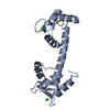
| Structure viewer | Molecule: MolmilJmol/JSmol |
|---|
- Downloads & links
-Download
| PDBx/mmCIF format | 6spv.cif.gz | 86.4 KB | Display | PDBx/mmCIF format |
|---|---|---|---|---|
| PDB format | pdb6spv.ent.gz | 63.4 KB | Display | PDB format |
| PDBx/mmJSON format | 6spv.json.gz | Tree view | PDBx/mmJSON format | |
| Others |  Other downloads Other downloads |
-Validation report
| Arichive directory | https://data.pdbj.org/pub/pdb/validation_reports/sp/6spvftp://data.pdbj.org/pub/pdb/validation_reports/sp/6spv | HTTPS FTP |
|---|
-Related structure data
| Related structure data | 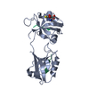 6spzC 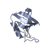 3rl7S 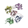 3rl8S S: Starting model for refinement C: citing same article ( |
|---|---|
| Similar structure data |


-Links
 PDBj
PDBj


- Assembly
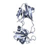
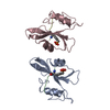
| Deposited unit | 
| |||||||||
|---|---|---|---|---|---|---|---|---|---|---|
| 1 |
| |||||||||
| Unit cell |
| |||||||||
| Components on special symmetry positions |
|
-Components
| #1: Protein | Mass: 20868.662 Da / Num. of mol.: 1 Source method: isolated from a genetically manipulated source Source: (gene. exp.) Homo sapiens (human) / Gene: DLG4, PSD95 / Plasmid: pOPINF / Production host:  |
|---|---|
| #2: Chemical | ChemComp-GSH /   Mass: 307.323 Da / Num. of mol.: 1 / Source method: obtained synthetically / Formula: C10H17N3O6S / Feature type: SUBJECT OF INVESTIGATION Mass: 307.323 Da / Num. of mol.: 1 / Source method: obtained synthetically / Formula: C10H17N3O6S / Feature type: SUBJECT OF INVESTIGATION |
| #3: Water | ChemComp-HOH /  Mass: 18.015 Da / Num. of mol.: 40 / Source method: isolated from a natural source / Formula: H2O Mass: 18.015 Da / Num. of mol.: 40 / Source method: isolated from a natural source / Formula: H2O |
| Has ligand of interest | Y |
-Experimental details
-Experiment
| Experiment | Method: X-RAY DIFFRACTION / Number of used crystals: 1 |
|---|
- Sample preparation
| Crystal | Density Matthews: 2.73 Å3/Da / Density % sol: 54.86 % / Description: Bi-pyramid |
|---|---|
| Crystal grow | Temperature: 277 K / Method: vapor diffusion, sitting drop / pH: 6.5 Details: 0.2 M calcium acetate; 0.1 M sodium cacodylate; 40% v/v PEG 400 |
-Data collection
| Diffraction | Mean temperature: 80 K / Serial crystal experiment: N |
|---|---|
| Diffraction source | Source: SYNCHROTRON / Site: Diamond  / Beamline: I04 / Wavelength: 0.92 Å / Beamline: I04 / Wavelength: 0.92 Å |
| Detector | Type: DECTRIS PILATUS 2M / Detector: PIXEL / Date: Jul 15, 2014 |
| Radiation | Protocol: SINGLE WAVELENGTH / Monochromatic (M) / Laue (L): M / Scattering type: x-ray |
| Radiation wavelength | Wavelength: 0.92 Å / Relative weight: 1 |
| Reflection | Resolution: 2.04→48.48 Å / Num. obs: 14015 / % possible obs: 99.4 % / Redundancy: 11.1 % / CC1/2: 0.999 / Rmerge(I) obs: 0.05 / Net I/σ(I): 24.4 |
| Reflection shell | Resolution: 2.04→2.09 Å / Redundancy: 5.5 % / Rmerge(I) obs: 0.547 / Mean I/σ(I) obs: 2.8 / Num. unique obs: 5594 / CC1/2: 0.851 / % possible all: 97.6 |
- Processing
| Software |
| ||||||||||||||||||||||||||||||||||||||||||||||||||||||||||||||||||||||||||||||||||||||||||||||||||||||||||||||||||||||||||||||||||||||||||||||||||||||||||||||||||||||||
|---|---|---|---|---|---|---|---|---|---|---|---|---|---|---|---|---|---|---|---|---|---|---|---|---|---|---|---|---|---|---|---|---|---|---|---|---|---|---|---|---|---|---|---|---|---|---|---|---|---|---|---|---|---|---|---|---|---|---|---|---|---|---|---|---|---|---|---|---|---|---|---|---|---|---|---|---|---|---|---|---|---|---|---|---|---|---|---|---|---|---|---|---|---|---|---|---|---|---|---|---|---|---|---|---|---|---|---|---|---|---|---|---|---|---|---|---|---|---|---|---|---|---|---|---|---|---|---|---|---|---|---|---|---|---|---|---|---|---|---|---|---|---|---|---|---|---|---|---|---|---|---|---|---|---|---|---|---|---|---|---|---|---|---|---|---|---|---|---|---|
| Refinement | Method to determine structure: MOLECULAR REPLACEMENT Starting model: 3rl7, 3rl8 Resolution: 2.04→48.48 Å / Cor.coef. Fo:Fc: 0.952 / Cor.coef. Fo:Fc free: 0.932 / WRfactor Rfree: 0.28 / WRfactor Rwork: 0.237 / SU B: 14.136 / SU ML: 0.206 / Cross valid method: FREE R-VALUE / ESU R: 0.219 / ESU R Free: 0.185 Details: Hydrogens have been added in their riding positions
| ||||||||||||||||||||||||||||||||||||||||||||||||||||||||||||||||||||||||||||||||||||||||||||||||||||||||||||||||||||||||||||||||||||||||||||||||||||||||||||||||||||||||
| Solvent computation | Ion probe radii: 0.8 Å / Shrinkage radii: 0.8 Å / VDW probe radii: 1.4 Å / Solvent model: BABINET MODEL PLUS MASK | ||||||||||||||||||||||||||||||||||||||||||||||||||||||||||||||||||||||||||||||||||||||||||||||||||||||||||||||||||||||||||||||||||||||||||||||||||||||||||||||||||||||||
| Displacement parameters | Biso mean: 65.303 Å2
| ||||||||||||||||||||||||||||||||||||||||||||||||||||||||||||||||||||||||||||||||||||||||||||||||||||||||||||||||||||||||||||||||||||||||||||||||||||||||||||||||||||||||
| Refinement step | Cycle: LAST / Resolution: 2.04→48.48 Å
| ||||||||||||||||||||||||||||||||||||||||||||||||||||||||||||||||||||||||||||||||||||||||||||||||||||||||||||||||||||||||||||||||||||||||||||||||||||||||||||||||||||||||
| Refine LS restraints |
| ||||||||||||||||||||||||||||||||||||||||||||||||||||||||||||||||||||||||||||||||||||||||||||||||||||||||||||||||||||||||||||||||||||||||||||||||||||||||||||||||||||||||
| LS refinement shell | Refine-ID: X-RAY DIFFRACTION / Total num. of bins used: 20
| ||||||||||||||||||||||||||||||||||||||||||||||||||||||||||||||||||||||||||||||||||||||||||||||||||||||||||||||||||||||||||||||||||||||||||||||||||||||||||||||||||||||||
| Refinement TLS params. | Method: refined / Refine-ID: X-RAY DIFFRACTION
| ||||||||||||||||||||||||||||||||||||||||||||||||||||||||||||||||||||||||||||||||||||||||||||||||||||||||||||||||||||||||||||||||||||||||||||||||||||||||||||||||||||||||
| Refinement TLS group |
|
