 Movie
Movie Controller


+ Open data

- Basic information
| Entry | Database: PDB / ID: 9rjc | |||||||||||||||||||||||||||
|---|---|---|---|---|---|---|---|---|---|---|---|---|---|---|---|---|---|---|---|---|---|---|---|---|---|---|---|---|
| Title | Apo Structure of the Human Signal Peptidase | |||||||||||||||||||||||||||
 Components Components |
| |||||||||||||||||||||||||||
 Keywords Keywords | MEMBRANE PROTEIN / Signal Peptidase Complex / protein biogenesis / secretory pathway / membrane thinning | |||||||||||||||||||||||||||
| Function / homology |  Function and homology information Function and homology informationsignal peptidase complex / signal peptidase I / signal peptidase activity / : / protein targeting to ER / Synthesis, secretion, and inactivation of Glucose-dependent Insulinotropic Polypeptide (GIP) / virion assembly / Regulation of CDH1 posttranslational processing and trafficking to plasma membrane / Maturation of hRSV A proteins / Synthesis, secretion, and deacylation of Ghrelin ...signal peptidase complex / signal peptidase I / signal peptidase activity / : / protein targeting to ER / Synthesis, secretion, and inactivation of Glucose-dependent Insulinotropic Polypeptide (GIP) / virion assembly / Regulation of CDH1 posttranslational processing and trafficking to plasma membrane / Maturation of hRSV A proteins / Synthesis, secretion, and deacylation of Ghrelin / SRP-dependent cotranslational protein targeting to membrane / Maturation of DENV proteins / response to virus / Synthesis, secretion, and inactivation of Glucagon-like Peptide-1 (GLP-1) / peptidase activity / viral protein processing / serine-type endopeptidase activity / endoplasmic reticulum membrane / endoplasmic reticulum / proteolysis Similarity search - Function | |||||||||||||||||||||||||||
| Biological species |  Homo sapiens (human) Homo sapiens (human) | |||||||||||||||||||||||||||
| Method | ELECTRON MICROSCOPY / single particle reconstruction / cryo EM / Resolution: 4.2 Å | |||||||||||||||||||||||||||
 Authors Authors | Liaci, A.M. / Vismpas, D. / Skalidis, I. / Koh, F.A. / Abhay, K. / Forster, G.F. | |||||||||||||||||||||||||||
| Funding support |  Netherlands, 2items Netherlands, 2items
| |||||||||||||||||||||||||||
 Citation Citation | Journal: Nat Commun / Year: 2026 Title: Structural basis of signal peptide recognition by the signal peptidase complex. Authors: A Manuel Liaci / Dimitrios Vismpas / Lisbeth R Kjølbye / Ioannis Skalidis / Gilberto P Pereira / Adrian Fujiet Koh / Mariska Gröllers-Mulderij / Abhay Kotecha / Paulo C T Souza / Friedrich Förster /   Abstract: The signal peptidase complex (SPC) is responsible for cleaving signal peptides (SPs) from approximately 10% of the human proteome. SPs are characterized by a tripartite structure, consisting of an N- ...The signal peptidase complex (SPC) is responsible for cleaving signal peptides (SPs) from approximately 10% of the human proteome. SPs are characterized by a tripartite structure, consisting of an N-terminal n-region, a central helical h-region and a C-terminal c-region, each defined by rather general chemical properties rather than strict sequence conservation. Despite their sequence diversity, SPC recognizes and processes SPs with exquisite specificity. Here, we present a 2.6 Å cryo-EM map of the human SPC-A, one of two SPC paralogs, bound to a model SP. The c-region binds to a hydrophobic binding groove near the active site, a narrow gate marks the transition from c- to h-region, and the h-region localizes in a transmembrane (TM) window. Substrate engagement stabilizes N- and C-terminal helices of Sec11A, which frame the SP and are unresolved in the apo structure. Molecular dynamics (MD) simulations confirm a stable hydrogen-bonding network at the c-region and indicate dynamic interactions within a thinned lipid environment at the TM window. AlphaFold modeling supports this binding mode across physiological SPs. Collectively, our structural and computational analyses explain how the SPC achieves its specificity by combining the selectivity of the luminal binding groove and of the transmembrane window. | |||||||||||||||||||||||||||
| History |
|
- Structure visualization
| Structure viewer | Molecule: MolmilJmol/JSmol |
|---|
- Downloads & links
-Download
| PDBx/mmCIF format | 9rjc.cif.gz | 108.5 KB | Display | PDBx/mmCIF format |
|---|---|---|---|---|
| PDB format | pdb9rjc.ent.gz | Display | PDB format | |
| PDBx/mmJSON format | 9rjc.json.gz | Tree view | PDBx/mmJSON format | |
| Others |  Other downloads Other downloads |
-Validation report
| Arichive directory | https://data.pdbj.org/pub/pdb/validation_reports/rj/9rjcftp://data.pdbj.org/pub/pdb/validation_reports/rj/9rjc | HTTPS FTP |
|---|
-Related structure data
| Related structure data |  54011MC  9rjbC M: map data used to model this data C: citing same article ( |
|---|---|
| Similar structure data |

-Links
 PDBj
PDBj





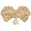
- Assembly
| Deposited unit | 
|
|---|---|
| 1 |
|
-Components
| #1: Protein | Mass: 20394.979 Da / Num. of mol.: 1 Source method: isolated from a genetically manipulated source Source: (gene. exp.) Homo sapiens (human) / Gene: SEC11A, SEC11L1, SPC18, SPCS4A / Production host: Homo sapiens (human) / References: UniProt: P67812, signal peptidase I |
|---|---|
| #2: Protein | Mass: 7386.763 Da / Num. of mol.: 1 Source method: isolated from a genetically manipulated source Source: (gene. exp.) Homo sapiens (human) / Gene: SPCS1, SPC12, HSPC033 / Production host: Homo sapiens (human) / References: UniProt: Q9Y6A9 |
| #3: Protein | Mass: 20338.516 Da / Num. of mol.: 1 Source method: isolated from a genetically manipulated source Source: (gene. exp.) Homo sapiens (human) / Gene: SPCS2, KIAA0102, SPC25 / Production host: Homo sapiens (human) / References: UniProt: Q15005 |
| #4: Protein | Mass: 19611.531 Da / Num. of mol.: 1 Source method: isolated from a genetically manipulated source Source: (gene. exp.) Homo sapiens (human) / Gene: SPCS3, SPC22, UNQ1841/PRO3567 / Production host: Homo sapiens (human) / References: UniProt: P61009 |
| Has protein modification | N |
-Experimental details
-Experiment
| Experiment | Method: ELECTRON MICROSCOPY |
|---|---|
| EM experiment | Aggregation state: PARTICLE / 3D reconstruction method: single particle reconstruction |
- Sample preparation
| Component | Name: Apo Structure of the Human Signal Peptidase Complex / Type: COMPLEX / Entity ID: #3, #1, #4, #2 / Source: RECOMBINANT | |||||||||||||||||||||||||
|---|---|---|---|---|---|---|---|---|---|---|---|---|---|---|---|---|---|---|---|---|---|---|---|---|---|---|
| Molecular weight | Value: 0.09334 MDa / Experimental value: NO | |||||||||||||||||||||||||
| Source (natural) | Organism: Homo sapiens (human) | |||||||||||||||||||||||||
| Source (recombinant) | Organism: Homo sapiens (human) / Cell: HEK 293 | |||||||||||||||||||||||||
| Buffer solution | pH: 7.8 Details: addition of 1.5 mM fluorinated, fos-choline (FFosC, Anatrace) directly before vitrification | |||||||||||||||||||||||||
| Buffer component |
| |||||||||||||||||||||||||
| Specimen | Conc.: 4 mg/ml / Embedding applied: NO / Shadowing applied: NO / Staining applied: NO / Vitrification applied: YES | |||||||||||||||||||||||||
| Specimen support | Grid material: COPPER / Grid mesh size: 200 divisions/in. / Grid type: Quantifoil Active R2/1 | |||||||||||||||||||||||||
| Vitrification | Instrument: FEI VITROBOT MARK IV / Cryogen name: ETHANE-PROPANE / Humidity: 100 % / Chamber temperature: 277 K |
- Electron microscopy imaging
| Experimental equipment |  Model: Titan Krios / Image courtesy: FEI Company |
|---|---|
| Microscopy | Model: TFS KRIOS |
| Electron gun | Electron source:  FIELD EMISSION GUN / Accelerating voltage: 300 kV / Illumination mode: FLOOD BEAM FIELD EMISSION GUN / Accelerating voltage: 300 kV / Illumination mode: FLOOD BEAM |
| Electron lens | Mode: BRIGHT FIELD / Nominal defocus max: 1200 nm / Nominal defocus min: 800 nm / Alignment procedure: COMA FREE |
| Image recording | Electron dose: 60 e/Å2 / Film or detector model: FEI FALCON IV (4k x 4k) / Num. of grids imaged: 1 / Num. of real images: 6537 |
- Processing
| EM software |
| ||||||||||||||||||||||||||||||||||||||||||||||||||
|---|---|---|---|---|---|---|---|---|---|---|---|---|---|---|---|---|---|---|---|---|---|---|---|---|---|---|---|---|---|---|---|---|---|---|---|---|---|---|---|---|---|---|---|---|---|---|---|---|---|---|---|
| CTF correction | Type: PHASE FLIPPING AND AMPLITUDE CORRECTION | ||||||||||||||||||||||||||||||||||||||||||||||||||
| Particle selection | Num. of particles selected: 2457714 | ||||||||||||||||||||||||||||||||||||||||||||||||||
| 3D reconstruction | Resolution: 4.2 Å / Resolution method: FSC 0.143 CUT-OFF / Num. of particles: 516615 / Algorithm: SIMULTANEOUS ITERATIVE (SIRT) / Num. of class averages: 1 / Symmetry type: POINT | ||||||||||||||||||||||||||||||||||||||||||||||||||
| Atomic model building | Protocol: FLEXIBLE FIT / Space: REAL | ||||||||||||||||||||||||||||||||||||||||||||||||||
| Atomic model building | PDB-ID: 7P2P Accession code: 7P2P / Details: PDB / Source name: PDB / Type: experimental model |

