 Movie
Movie Controller


+ Open data

- Basic information
| Entry | Database: PDB / ID: 7p2p | ||||||
|---|---|---|---|---|---|---|---|
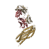
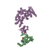
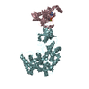
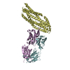
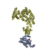
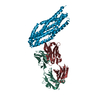
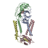
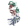
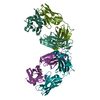
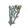
| Title | Human Signal Peptidase Complex Paralog A (SPC-A) | ||||||
 Components Components |
| ||||||
 Keywords Keywords | MEMBRANE PROTEIN / Endoplasmic Reticulum / Signal peptide / Serine protease / Membrane complex | ||||||
| Function / homology |  Function and homology information Function and homology informationsignal peptidase complex / signal peptidase I / signal peptidase activity / : / protein targeting to ER / Synthesis, secretion, and inactivation of Glucose-dependent Insulinotropic Polypeptide (GIP) / virion assembly / Regulation of CDH1 posttranslational processing and trafficking to plasma membrane / Maturation of hRSV A proteins / Synthesis, secretion, and deacylation of Ghrelin ...signal peptidase complex / signal peptidase I / signal peptidase activity / : / protein targeting to ER / Synthesis, secretion, and inactivation of Glucose-dependent Insulinotropic Polypeptide (GIP) / virion assembly / Regulation of CDH1 posttranslational processing and trafficking to plasma membrane / Maturation of hRSV A proteins / Synthesis, secretion, and deacylation of Ghrelin / SRP-dependent cotranslational protein targeting to membrane / response to virus / Synthesis, secretion, and inactivation of Glucagon-like Peptide-1 (GLP-1) / peptidase activity / viral protein processing / serine-type endopeptidase activity / endoplasmic reticulum membrane / endoplasmic reticulum / proteolysis Similarity search - Function | ||||||
| Biological species |  Homo sapiens (human) Homo sapiens (human) | ||||||
| Method | ELECTRON MICROSCOPY / single particle reconstruction / cryo EM / Resolution: 4.9 Å | ||||||
 Authors Authors | Liaci, A.M. / Foerster, F. | ||||||
| Funding support |  Netherlands, 1items Netherlands, 1items
| ||||||
 Citation Citation | Journal: Mol Cell / Year: 2021 Title: Structure of the human signal peptidase complex reveals the determinants for signal peptide cleavage. Authors: A Manuel Liaci / Barbara Steigenberger / Paulo Cesar Telles de Souza / Sem Tamara / Mariska Gröllers-Mulderij / Patrick Ogrissek / Siewert J Marrink / Richard A Scheltema / Friedrich Förster /   Abstract: The signal peptidase complex (SPC) is an essential membrane complex in the endoplasmic reticulum (ER), where it removes signal peptides (SPs) from a large variety of secretory pre-proteins with ...The signal peptidase complex (SPC) is an essential membrane complex in the endoplasmic reticulum (ER), where it removes signal peptides (SPs) from a large variety of secretory pre-proteins with exquisite specificity. Although the determinants of this process have been established empirically, the molecular details of SP recognition and removal remain elusive. Here, we show that the human SPC exists in two functional paralogs with distinct proteolytic subunits. We determined the atomic structures of both paralogs using electron cryo-microscopy and structural proteomics. The active site is formed by a catalytic triad and abuts the ER membrane, where a transmembrane window collectively formed by all subunits locally thins the bilayer. Molecular dynamics simulations indicate that this unique architecture generates specificity for SPs based on the length of their hydrophobic segments. #1: Journal: BioRxiv / Year: 2020Title: Structure of the Human Signal Peptidase Complex Reveals the Determinants for Signal Peptide Cleavage Authors: Liaci, A.M. / Steigenberger, B. / Tamara, S. / Telles de Souza, P.C. / Grollers-Mulderij, M. / Ogrissek, P. / Marrik, S.J. / Scheltema, R.A. / Foerster, F. | ||||||
| History |
|
- Structure visualization
| Movie |
Movie viewer |
|---|---|
| Structure viewer | Molecule: MolmilJmol/JSmol |
- Downloads & links
-Download
| PDBx/mmCIF format | 7p2p.cif.gz | 127.3 KB | Display | PDBx/mmCIF format |
|---|---|---|---|---|
| PDB format | pdb7p2p.ent.gz | 93.3 KB | Display | PDB format |
| PDBx/mmJSON format | 7p2p.json.gz | Tree view | PDBx/mmJSON format | |
| Others |  Other downloads Other downloads |
-Validation report
| Arichive directory | https://data.pdbj.org/pub/pdb/validation_reports/p2/7p2pftp://data.pdbj.org/pub/pdb/validation_reports/p2/7p2p | HTTPS FTP |
|---|
-Related structure data
| Related structure data | 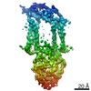 13171MC  7p2qC C: citing same article ( M: map data used to model this data |
|---|---|
| Similar structure data |

-Links
 PDBj
PDBj


- Assembly
| Deposited unit | 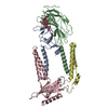
|
|---|---|
| 1 |
|
-Components
| #1: Protein | Mass: 22376.088 Da / Num. of mol.: 1 Source method: isolated from a genetically manipulated source Source: (gene. exp.) Homo sapiens (human) / Gene: SEC11A, SEC11L1, SPC18, SPCS4A / Plasmid: pUPE-2961 / Cell line (production host): HEK 293 E+ / Production host: Homo sapiens (human) / References: UniProt: P67812, signal peptidase I |
|---|---|
| #2: Protein | Mass: 22348.457 Da / Num. of mol.: 1 Source method: isolated from a genetically manipulated source Source: (gene. exp.) Homo sapiens (human) / Gene: SPCS3, SPC22, UNQ1841/PRO3567 / Plasmid: pUPE-2961 / Cell line (production host): HEK 293 E+ / Production host: Homo sapiens (human)References: UniProt: P61009, Hydrolases; Acting on peptide bonds (peptidases) |
| #3: Protein | Mass: 27215.236 Da / Num. of mol.: 1 Source method: isolated from a genetically manipulated source Source: (gene. exp.) Homo sapiens (human) / Gene: SPCS2, KIAA0102, SPC25 / Plasmid: pUPE-2961 / Cell line (production host): HEK 293 E+ / Production host: Homo sapiens (human)References: UniProt: Q15005, Hydrolases; Acting on peptide bonds (peptidases) |
| #4: Protein | Mass: 20488.445 Da / Num. of mol.: 1 Source method: isolated from a genetically manipulated source Source: (gene. exp.) Homo sapiens (human) / Gene: SPCS1, SPC12, HSPC033 / Plasmid: pUPE-2961 / Cell line (production host): HEK 293 E+ / Production host: Homo sapiens (human)References: UniProt: Q9Y6A9, Hydrolases; Acting on peptide bonds (peptidases) |
| #5: Polysaccharide | alpha-D-mannopyranose-(1-3)-[alpha-D-mannopyranose-(1-6)]beta-D-mannopyranose-(1-4)-2-acetamido-2- ...alpha-D-mannopyranose-(1-3)-[alpha-D-mannopyranose-(1-6)]beta-D-mannopyranose-(1-4)-2-acetamido-2-deoxy-beta-D-glucopyranose-(1-4)-2-acetamido-2-deoxy-beta-D-glucopyranose Source method: isolated from a genetically manipulated source |
| Has ligand of interest | N |
| Has protein modification | Y |
-Experimental details
-Experiment
| Experiment | Method: ELECTRON MICROSCOPY |
|---|---|
| EM experiment | Aggregation state: PARTICLE / 3D reconstruction method: single particle reconstruction |
- Sample preparation
| Component |
| ||||||||||||||||||||||||||||||||||||||||||||||||||
|---|---|---|---|---|---|---|---|---|---|---|---|---|---|---|---|---|---|---|---|---|---|---|---|---|---|---|---|---|---|---|---|---|---|---|---|---|---|---|---|---|---|---|---|---|---|---|---|---|---|---|---|
| Molecular weight |
| ||||||||||||||||||||||||||||||||||||||||||||||||||
| Source (natural) |
| ||||||||||||||||||||||||||||||||||||||||||||||||||
| Source (recombinant) |
| ||||||||||||||||||||||||||||||||||||||||||||||||||
| Buffer solution |
| ||||||||||||||||||||||||||||||||||||||||||||||||||
| Buffer component |
| ||||||||||||||||||||||||||||||||||||||||||||||||||
| Specimen | Experiment-ID: 1 / Embedding applied: NO / Shadowing applied: NO / Staining applied: NO / Vitrification applied: YES
| ||||||||||||||||||||||||||||||||||||||||||||||||||
| Specimen support |
| ||||||||||||||||||||||||||||||||||||||||||||||||||
| Vitrification | Chamber temperature: 277 K / Cryogen name: ETHANE / Details: Blot for 4s with blot force 0 before plunging. / Entry-ID: 7P2P / Humidity: 100 % / Instrument: FEI VITROBOT MARK IV
|
- Electron microscopy imaging
| Experimental equipment |  Model: Talos Arctica / Image courtesy: FEI Company | |||||||||
|---|---|---|---|---|---|---|---|---|---|---|
| Microscopy | Model: FEI TALOS ARCTICA Details: 200 kV Talos Arctica at Utrecht University, the Netherlands. Same settings were used for both grid conditions. | |||||||||
| Electron gun | Electron source:  FIELD EMISSION GUN / Accelerating voltage: 200 kV / Illumination mode: FLOOD BEAM FIELD EMISSION GUN / Accelerating voltage: 200 kV / Illumination mode: FLOOD BEAM | |||||||||
| Electron lens | Mode: BRIGHT FIELD / Nominal magnification: 165000 X / Nominal defocus max: 4000 nm / Nominal defocus min: 1000 nm / Calibrated defocus min: 300 nm / Calibrated defocus max: 4800 nm / Cs: 2.7 mm / C2 aperture diameter: 30 µm / Alignment procedure: COMA FREE | |||||||||
| Specimen holder | Cryogen: NITROGEN / Specimen holder model: FEI TITAN KRIOS AUTOGRID HOLDER / Temperature (max): 93 K / Temperature (min): 93 K | |||||||||
| Image recording | Imaging-ID: 1 / Electron dose: 63 e/Å2 / Detector mode: COUNTING / Film or detector model: GATAN K2 SUMMIT (4k x 4k) / Num. of grids imaged: 1
| |||||||||
| EM imaging optics | Energyfilter name: GIF Quantum LS / Energyfilter slit width: 20 eV | |||||||||
| Image scans | Width: 3838 / Height: 3710 / Movie frames/image: 51 |
- Processing
| Software | Name: PHENIX / Version: 1.13_2998: / Classification: refinement | ||||||||||||||||||||||||||||||||||||||||
|---|---|---|---|---|---|---|---|---|---|---|---|---|---|---|---|---|---|---|---|---|---|---|---|---|---|---|---|---|---|---|---|---|---|---|---|---|---|---|---|---|---|
| EM software |
| ||||||||||||||||||||||||||||||||||||||||
| CTF correction | Type: PHASE FLIPPING AND AMPLITUDE CORRECTION | ||||||||||||||||||||||||||||||||||||||||
| Particle selection | Num. of particles selected: 1015857 | ||||||||||||||||||||||||||||||||||||||||
| 3D reconstruction | Resolution: 4.9 Å / Resolution method: FSC 0.143 CUT-OFF / Num. of particles: 29508 / Num. of class averages: 1 / Symmetry type: POINT | ||||||||||||||||||||||||||||||||||||||||
| Atomic model building | Protocol: AB INITIO MODEL / Space: REAL |
