- EMDB-54010: Human Signal Peptidase in complex with artificial Signal Peptide L11 -
+
Open data
ID or keywords:
Loading...
-
Basic information
Entry
Database: EMDB / ID: EMD-54010
Title
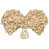
Human Signal Peptidase in complex with artificial Signal Peptide L11
Map data
Sample
Complex: Signal peptidase complex with a signal peptide
Protein or peptide: Signal peptidase complex subunit 2
Protein or peptide: Signal peptidase complex catalytic subunit SEC11A
Protein or peptide: Signal peptidase complex subunit 3
Protein or peptide: Signal peptidase complex subunit 1
Protein or peptide: Artificial Signal Peptide L11
Keywords
Signal Peptidase Complex / Signal Peptide / protein biogenesis / secretory pathway / membrane thinning / MEMBRANE PROTEIN
Function / homology
Function and homology information
signal peptidase complex / signal peptidase I / signal peptidase activity / : / protein targeting to ER / Synthesis, secretion, and inactivation of Glucose-dependent Insulinotropic Polypeptide (GIP) / virion assembly / Regulation of CDH1 posttranslational processing and trafficking to plasma membrane / Maturation of hRSV A proteins / Synthesis, secretion, and deacylation of Ghrelin ...signal peptidase complex / signal peptidase I / signal peptidase activity / : / protein targeting to ER / Synthesis, secretion, and inactivation of Glucose-dependent Insulinotropic Polypeptide (GIP) / virion assembly / Regulation of CDH1 posttranslational processing and trafficking to plasma membrane / Maturation of hRSV A proteins / Synthesis, secretion, and deacylation of Ghrelin / SRP-dependent cotranslational protein targeting to membrane / Maturation of DENV proteins / response to virus / Synthesis, secretion, and inactivation of Glucagon-like Peptide-1 (GLP-1) / peptidase activity / viral protein processing / serine-type endopeptidase activity / endoplasmic reticulum membrane / endoplasmic reticulum / proteolysis Similarity search - Function
Peptidase S26B / Signal peptidase complex subunit 3 / Signal peptidase complex subunit 2 / Signal peptidase subunit / Microsomal signal peptidase 25 kDa subunit (SPC25) / Signal peptidase complex subunit 1 / Microsomal signal peptidase 12 kDa subunit (SPC12) / Peptidase S26A, signal peptidase I, conserved site / Signal peptidases I signature 3. / Peptidase S26A, signal peptidase I, serine active site ...Peptidase S26B / Signal peptidase complex subunit 3 / Signal peptidase complex subunit 2 / Signal peptidase subunit / Microsomal signal peptidase 25 kDa subunit (SPC25) / Signal peptidase complex subunit 1 / Microsomal signal peptidase 12 kDa subunit (SPC12) / Peptidase S26A, signal peptidase I, conserved site / Signal peptidases I signature 3. / Peptidase S26A, signal peptidase I, serine active site / Signal peptidases I serine active site. / Peptidase S26 / Peptidase S24/S26A/S26B/S26C / Peptidase S24-like / LexA/Signal peptidase-like superfamily Similarity search - Domain/homology
Signal peptidase complex subunit 3 / Signal peptidase complex catalytic subunit SEC11A / Signal peptidase complex subunit 2 / Signal peptidase complex subunit 1 Similarity search - Component
Biological species
Homo sapiens (human)
Method
single particle reconstruction / cryo EM / Resolution: 2.6 Å
Netherlands Organisation for Scientific Research (NWO)
OCENW.M.22.151
Netherlands
Netherlands Organisation for Scientific Research (NWO)
184.034.014
Netherlands
Citation
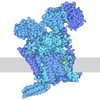
Journal: Nat Commun / Year: 2026 Title: Structural basis of signal peptide recognition by the signal peptidase complex. Authors: A Manuel Liaci / Dimitrios Vismpas / Lisbeth R Kjølbye / Ioannis Skalidis / Gilberto P Pereira / Adrian Fujiet Koh / Mariska Gröllers-Mulderij / Abhay Kotecha / Paulo C T Souza / Friedrich Förster / Abstract: The signal peptidase complex (SPC) is responsible for cleaving signal peptides (SPs) from approximately 10% of the human proteome. SPs are characterized by a tripartite structure, consisting of an N- ...The signal peptidase complex (SPC) is responsible for cleaving signal peptides (SPs) from approximately 10% of the human proteome. SPs are characterized by a tripartite structure, consisting of an N-terminal n-region, a central helical h-region and a C-terminal c-region, each defined by rather general chemical properties rather than strict sequence conservation. Despite their sequence diversity, SPC recognizes and processes SPs with exquisite specificity. Here, we present a 2.6 Å cryo-EM map of the human SPC-A, one of two SPC paralogs, bound to a model SP. The c-region binds to a hydrophobic binding groove near the active site, a narrow gate marks the transition from c- to h-region, and the h-region localizes in a transmembrane (TM) window. Substrate engagement stabilizes N- and C-terminal helices of Sec11A, which frame the SP and are unresolved in the apo structure. Molecular dynamics (MD) simulations confirm a stable hydrogen-bonding network at the c-region and indicate dynamic interactions within a thinned lipid environment at the TM window. AlphaFold modeling supports this binding mode across physiological SPs. Collectively, our structural and computational analyses explain how the SPC achieves its specificity by combining the selectivity of the luminal binding groove and of the transmembrane window.
In the structure databanks used in Yorodumi, some data are registered as the other names, "COVID-19 virus" and "2019-nCoV". Here are the details of the virus and the list of structure data.
Jan 31, 2019. EMDB accession codes are about to change! (news from PDBe EMDB page)
EMDB accession codes are about to change! (news from PDBe EMDB page)
The allocation of 4 digits for EMDB accession codes will soon come to an end. Whilst these codes will remain in use, new EMDB accession codes will include an additional digit and will expand incrementally as the available range of codes is exhausted. The current 4-digit format prefixed with “EMD-” (i.e. EMD-XXXX) will advance to a 5-digit format (i.e. EMD-XXXXX), and so on. It is currently estimated that the 4-digit codes will be depleted around Spring 2019, at which point the 5-digit format will come into force.
The EM Navigator/Yorodumi systems omit the EMD- prefix.
Related info.:Q: What is EMD? / ID/Accession-code notation in Yorodumi/EM Navigator
Yorodumi is a browser for structure data from EMDB, PDB, SASBDB, etc.
This page is also the successor to EM Navigator detail page, and also detail information page/front-end page for Omokage search.
The word "yorodu" (or yorozu) is an old Japanese word meaning "ten thousand". "mi" (miru) is to see.
Related info.:EMDB / PDB / SASBDB / Comparison of 3 databanks / Yorodumi Search / Aug 31, 2016. New EM Navigator & Yorodumi / Yorodumi Papers / Jmol/JSmol / Function and homology information / Changes in new EM Navigator and Yorodumi
 Movie
Movie Controller

 Yorodumi
 Open data
 Basic information
 Map data
 Sample
 Keywords
 Function and homology information
 Homo sapiens (human)
 Authors
 Netherlands, 2 items
  Citation

 Structure visualization
 Downloads & links
 emd_54010.png
 http://ftp.pdbj.org/pub/emdb/structures/EMD-54010








 Z (Sec.)
 Y (Row.)
 X (Col.)


 Sample components
 Processing
 Electron microscopy
 FIELD EMISSION GUN


