Movie
Movie Controller

[English] 日本語
 Yorodumi
- PDB-9nkj: Structure of substrates-engaged MIDN-bound human 26S proteasome,E... -
+ Open data

- Basic information
| Entry | Database: PDB / ID: 9nkj | |||||||||||||||
|---|---|---|---|---|---|---|---|---|---|---|---|---|---|---|---|---|
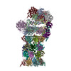
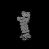
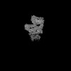
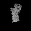
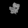
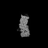
| Title | Structure of substrates-engaged MIDN-bound human 26S proteasome,ED-MIDN state (Composite map) | |||||||||||||||
 Components Components |
| |||||||||||||||
 Keywords Keywords | IMMUNE SYSTEM / Ubiquitin-indepedent / proteasome / Midnolin / 26S complex / ED-state | |||||||||||||||
| Function / homology |  Function and homology information Function and homology informationthyrotropin-releasing hormone receptor binding / nuclear proteasome complex / host-mediated perturbation of viral transcription / positive regulation of inclusion body assembly / Impaired BRCA2 translocation to the nucleus / Impaired BRCA2 binding to SEM1 (DSS1) / meiosis I / proteasome accessory complex / purine ribonucleoside triphosphate binding / integrator complex ...thyrotropin-releasing hormone receptor binding / nuclear proteasome complex / host-mediated perturbation of viral transcription / positive regulation of inclusion body assembly / Impaired BRCA2 translocation to the nucleus / Impaired BRCA2 binding to SEM1 (DSS1) / meiosis I / proteasome accessory complex / purine ribonucleoside triphosphate binding / integrator complex / proteasome regulatory particle / CD8-positive, alpha-beta T cell differentiation / thymic T cell selection / CD8-positive, alpha-beta T cell homeostasis / cytosolic proteasome complex / positive regulation of proteasomal protein catabolic process / proteasome-activating activity / Antigen processing: Ub, ATP-independent proteasomal degradation / proteasome regulatory particle, lid subcomplex / proteasome regulatory particle, base subcomplex / T-helper 1 cell differentiation / negative regulation of regulatory T cell differentiation / cellular response to type I interferon / protein K63-linked deubiquitination / metal-dependent deubiquitinase activity / negative regulation of programmed cell death / Regulation of ornithine decarboxylase (ODC) / proteasome core complex / Proteasome assembly / T-helper 17 cell differentiation / Cross-presentation of soluble exogenous antigens (endosomes) / transcription factor binding / K63-linked deubiquitinase activity / Somitogenesis / Homologous DNA Pairing and Strand Exchange / Defective homologous recombination repair (HRR) due to BRCA1 loss of function / Defective HDR through Homologous Recombination Repair (HRR) due to PALB2 loss of BRCA1 binding function / Defective HDR through Homologous Recombination Repair (HRR) due to PALB2 loss of BRCA2/RAD51/RAD51C binding function / Resolution of D-loop Structures through Synthesis-Dependent Strand Annealing (SDSA) / flagellated sperm motility / Resolution of D-loop Structures through Holliday Junction Intermediates / proteasome binding / Impaired BRCA2 binding to RAD51 / regulation of protein catabolic process / myofibril / AMPK-induced ERAD and lysosome mediated degradation of PD-L1(CD274) / GSK3B-mediated proteasomal degradation of PD-L1(CD274) / SPOP-mediated proteasomal degradation of PD-L1(CD274) / proteasomal ubiquitin-independent protein catabolic process / Ribosome Quality Control (RQC) complex extracts and degrades nascent peptide / general transcription initiation factor binding / proteasome storage granule / Presynaptic phase of homologous DNA pairing and strand exchange / protein deubiquitination / polyubiquitin modification-dependent protein binding / proteasome endopeptidase complex / NF-kappaB binding / proteasome core complex, beta-subunit complex / endopeptidase activator activity / threonine-type endopeptidase activity / mRNA export from nucleus / proteasome core complex, alpha-subunit complex / proteasome assembly / immune system process / regulation of G1/S transition of mitotic cell cycle / enzyme regulator activity / ciliary tip / response to type II interferon / positive regulation of interleukin-2 production / inclusion body / regulation of proteasomal protein catabolic process / TBP-class protein binding / : / proteasome complex / stem cell differentiation / sarcomere / proteasomal protein catabolic process / Regulation of activated PAK-2p34 by proteasome mediated degradation / sperm end piece / ubiquitin binding / Autodegradation of Cdh1 by Cdh1:APC/C / APC/C:Cdc20 mediated degradation of Securin / negative regulation of inflammatory response to antigenic stimulus / N-glycan trimming in the ER and Calnexin/Calreticulin cycle / Asymmetric localization of PCP proteins / Ubiquitin-dependent degradation of Cyclin D / lipopolysaccharide binding / SCF-beta-TrCP mediated degradation of Emi1 / NIK-->noncanonical NF-kB signaling / AUF1 (hnRNP D0) binds and destabilizes mRNA / TNFR2 non-canonical NF-kB pathway / Assembly of the pre-replicative complex / Vpu mediated degradation of CD4 / P-body / Cdc20:Phospho-APC/C mediated degradation of Cyclin A / Dectin-1 mediated noncanonical NF-kB signaling / Degradation of DVL / Degradation of AXIN / Degradation of CRY and PER proteins / meiotic cell cycle Similarity search - Function | |||||||||||||||
| Biological species |  Homo sapiens (human) Homo sapiens (human) | |||||||||||||||
| Method | ELECTRON MICROSCOPY / single particle reconstruction / cryo EM / Resolution: 3.84 Å | |||||||||||||||
 Authors Authors | Peddada, N. / Beutler, B. | |||||||||||||||
| Funding support |  United States, 2items United States, 2items
| |||||||||||||||
 Citation Citation | Journal: Proc Natl Acad Sci U S A / Year: 2025 Title: Structural insights into the ubiquitin-independent midnolin-proteasome pathway. Authors: Nagesh Peddada / Xue Zhong / Yan Yin / Danielle Renee Lazaro / Jianhui Wang / Stephen Lyon / Jin Huk Choi / Xiao-Chen Bai / Eva Marie Y Moresco / Bruce Beutler / Abstract: The protein midnolin (MIDN) augments proteasome activity in lymphocytes and dramatically facilitates the survival and proliferation of B-lymphoid malignancies. MIDN binds both to proteasomes and to ...The protein midnolin (MIDN) augments proteasome activity in lymphocytes and dramatically facilitates the survival and proliferation of B-lymphoid malignancies. MIDN binds both to proteasomes and to substrates, but the mode of interaction with the proteasome is unknown, and the mechanism by which MIDN facilitates substrate degradation in a ubiquitin-independent manner is incompletely understood. Here, we present cryoelectron microscopy (cryo-EM) structures of the substrate-engaged, MIDN-bound human proteasome in two conformational states. MIDN induces proteasome conformations similarly to ubiquitinated substrates by using its ubiquitin-like domain to bind to the deubiquitinase RPN11 (PSMD14). By simultaneously binding to RPN1 (PSMD2) with its C-terminal α-helix, MIDN positions its substrate-carrying Catch domain above the proteasome ATPase channel through which substrates are translocated before degradation. Our findings suggest that both ubiquitin-like domain and C-terminal α-helix must bind to the proteasome for MIDN to stimulate proteasome activity. | |||||||||||||||
| History |
|
- Structure visualization
| Structure viewer | Molecule: MolmilJmol/JSmol |
|---|
- Downloads & links
-Download
| PDBx/mmCIF format | 9nkj.cif.gz | 3.1 MB | Display | PDBx/mmCIF format |
|---|---|---|---|---|
| PDB format | pdb9nkj.ent.gz | Display | PDB format | |
| PDBx/mmJSON format | 9nkj.json.gz | Tree view | PDBx/mmJSON format | |
| Others |  Other downloads Other downloads |
-Validation report
| Arichive directory | https://data.pdbj.org/pub/pdb/validation_reports/nk/9nkjftp://data.pdbj.org/pub/pdb/validation_reports/nk/9nkj | HTTPS FTP |
|---|
-Related structure data
| Related structure data |  49510MC 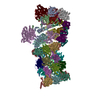 9nkfC  9nkgC 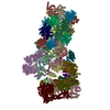 9nkiC C: citing same article ( M: map data used to model this data |
|---|---|
| Similar structure data |
-Links
 PDBj
PDBj


- Assembly
| Deposited unit | 
|
|---|---|
| 1 |
|
-Components
-26S proteasome regulatory subunit ... , 5 types, 5 molecules ABDEF
| #1: Protein | Mass: 48700.805 Da / Num. of mol.: 1 / Source method: isolated from a natural source / Source: (natural) Homo sapiens (human) / Cell line: HEK / References: UniProt: P35998 |
|---|---|
| #2: Protein | Mass: 49260.504 Da / Num. of mol.: 1 / Source method: isolated from a natural source / Source: (natural) Homo sapiens (human) / References: UniProt: P62191 |
| #4: Protein | Mass: 47426.141 Da / Num. of mol.: 1 / Source method: isolated from a natural source / Source: (natural) Homo sapiens (human) / Cell line: HEK / References: UniProt: P43686 |
| #5: Protein | Mass: 45867.027 Da / Num. of mol.: 1 / Source method: isolated from a natural source / Source: (natural) Homo sapiens (human) / Cell line: HEK / References: UniProt: A0A087X2I1 |
| #6: Protein | Mass: 49266.457 Da / Num. of mol.: 1 / Source method: isolated from a natural source / Source: (natural) Homo sapiens (human) / Cell line: HEK / References: UniProt: P17980 |
-Protein , 2 types, 2 molecules Ce
| #3: Protein | Mass: 45694.047 Da / Num. of mol.: 1 / Source method: isolated from a natural source / Source: (natural) Homo sapiens (human) / References: UniProt: P62195 |
|---|---|
| #19: Protein | Mass: 8284.611 Da / Num. of mol.: 1 / Source method: isolated from a natural source / Source: (natural) Homo sapiens (human) / Cell line: HEK / References: UniProt: P60896 |
-26S proteasome non-ATPase regulatory subunit ... , 11 types, 11 molecules UVXYZabcdfW
| #7: Protein | Mass: 105958.234 Da / Num. of mol.: 1 / Source method: isolated from a natural source / Source: (natural) Homo sapiens (human) / Cell line: HEK / References: UniProt: Q99460 |
|---|---|
| #8: Protein | Mass: 61066.500 Da / Num. of mol.: 1 / Source method: isolated from a natural source / Source: (natural) Homo sapiens (human) / References: UniProt: O43242 |
| #9: Protein | Mass: 47526.688 Da / Num. of mol.: 1 / Source method: isolated from a natural source / Source: (natural) Homo sapiens (human) / Cell line: HEK / References: UniProt: O00231 |
| #10: Protein | Mass: 45592.285 Da / Num. of mol.: 1 / Source method: isolated from a natural source / Source: (natural) Homo sapiens (human) / Cell line: HEK / References: UniProt: Q15008 |
| #11: Protein | Mass: 37086.441 Da / Num. of mol.: 1 / Source method: isolated from a natural source / Source: (natural) Homo sapiens (human) / Cell line: HEK / References: UniProt: P51665 |
| #12: Protein | Mass: 42995.359 Da / Num. of mol.: 1 / Source method: isolated from a natural source / Source: (natural) Homo sapiens (human) / Cell line: HEK / References: UniProt: Q9UNM6 |
| #13: Protein | Mass: 40781.590 Da / Num. of mol.: 1 / Source method: isolated from a natural source / Source: (natural) Homo sapiens (human) / Cell line: HEK / References: UniProt: P55036 |
| #14: Protein | Mass: 34620.023 Da / Num. of mol.: 1 / Source method: isolated from a natural source / Source: (natural) Homo sapiens (human) / Cell line: HEKReferences: UniProt: O00487, Hydrolases; Acting on peptide bonds (peptidases); Omega peptidases |
| #15: Protein | Mass: 39667.871 Da / Num. of mol.: 1 / Source method: isolated from a natural source / Source: (natural) Homo sapiens (human) / Cell line: HEK / References: UniProt: P48556 |
| #16: Protein | Mass: 100313.625 Da / Num. of mol.: 1 / Source method: isolated from a natural source / Source: (natural) Homo sapiens (human) / Cell line: HEK / References: UniProt: Q13200 |
| #18: Protein | Mass: 52979.359 Da / Num. of mol.: 1 / Source method: isolated from a natural source / Source: (natural) Homo sapiens (human) / Cell line: HEK / References: UniProt: O00232 |
-Protein/peptide , 1 types, 1 molecules v
| #17: Protein/peptide | Mass: 698.854 Da / Num. of mol.: 1 / Source method: isolated from a natural source / Details: its poly peptide- substrate density. / Source: (natural) Homo sapiens (human) / Cell line: HEK |
|---|
-Proteasome subunit alpha type- ... , 7 types, 14 molecules GgHhIiJjKkLlMm
| #20: Protein | Mass: 27432.459 Da / Num. of mol.: 2 / Source method: isolated from a natural source / Source: (natural) Homo sapiens (human) / Cell line: HEKReferences: UniProt: P60900, proteasome endopeptidase complex #21: Protein | Mass: 25927.535 Da / Num. of mol.: 2 / Source method: isolated from a natural source / Source: (natural) Homo sapiens (human) / Cell line: HEKReferences: UniProt: P25787, proteasome endopeptidase complex #22: Protein | Mass: 29525.842 Da / Num. of mol.: 2 / Source method: isolated from a natural source / Source: (natural) Homo sapiens (human) / Cell line: HEKReferences: UniProt: P25789, proteasome endopeptidase complex #23: Protein | Mass: 27929.891 Da / Num. of mol.: 2 / Source method: isolated from a natural source / Source: (natural) Homo sapiens (human) / References: UniProt: O14818#24: Protein | Mass: 26435.977 Da / Num. of mol.: 2 / Source method: isolated from a natural source / Source: (natural) Homo sapiens (human) / Cell line: HEKReferences: UniProt: P28066, proteasome endopeptidase complex #25: Protein | Mass: 29595.627 Da / Num. of mol.: 2 / Source method: isolated from a natural source / Source: (natural) Homo sapiens (human) / Cell line: HEKReferences: UniProt: P25786, proteasome endopeptidase complex #26: Protein | Mass: 28469.252 Da / Num. of mol.: 2 / Source method: isolated from a natural source / Source: (natural) Homo sapiens (human) / Cell line: HEKReferences: UniProt: P25788, proteasome endopeptidase complex |
|---|
-Proteasome subunit beta type- ... , 7 types, 14 molecules NnOoPpQqRrSsTt
| #27: Protein | Mass: 25377.652 Da / Num. of mol.: 2 / Source method: isolated from a natural source / Source: (natural) Homo sapiens (human) / Cell line: HEKReferences: UniProt: P28072, proteasome endopeptidase complex #28: Protein | Mass: 30000.418 Da / Num. of mol.: 2 / Source method: isolated from a natural source / Source: (natural) Homo sapiens (human) / Cell line: HEKReferences: UniProt: Q99436, proteasome endopeptidase complex #29: Protein | Mass: 22972.896 Da / Num. of mol.: 2 / Source method: isolated from a natural source / Source: (natural) Homo sapiens (human) / Cell line: HEKReferences: UniProt: P49720, proteasome endopeptidase complex #30: Protein | Mass: 22864.277 Da / Num. of mol.: 2 / Source method: isolated from a natural source / Source: (natural) Homo sapiens (human) / Cell line: HEKReferences: UniProt: P49721, proteasome endopeptidase complex #31: Protein | Mass: 28510.248 Da / Num. of mol.: 2 / Source method: isolated from a natural source / Source: (natural) Homo sapiens (human) / Cell line: HEKReferences: UniProt: P28074, proteasome endopeptidase complex #32: Protein | Mass: 26522.396 Da / Num. of mol.: 2 / Source method: isolated from a natural source / Source: (natural) Homo sapiens (human) / Cell line: HEKReferences: UniProt: P20618, proteasome endopeptidase complex #33: Protein | Mass: 29231.178 Da / Num. of mol.: 2 / Source method: isolated from a natural source / Source: (natural) Homo sapiens (human) / Cell line: HEKReferences: UniProt: P28070, proteasome endopeptidase complex |
|---|
-Non-polymers , 5 types, 15 molecules 


| #34: Chemical | ChemComp-ATP /  Mass: 507.181 Da / Num. of mol.: 4 / Source method: obtained synthetically / Formula: C10H16N5O13P3 / Feature type: SUBJECT OF INVESTIGATION / Comment: ATP, energy-carrying molecule*YM Mass: 507.181 Da / Num. of mol.: 4 / Source method: obtained synthetically / Formula: C10H16N5O13P3 / Feature type: SUBJECT OF INVESTIGATION / Comment: ATP, energy-carrying molecule*YM#35: Chemical |  Mass: 24.305 Da / Num. of mol.: 3 / Source method: obtained synthetically / Formula: Mg / Feature type: SUBJECT OF INVESTIGATION Mass: 24.305 Da / Num. of mol.: 3 / Source method: obtained synthetically / Formula: Mg / Feature type: SUBJECT OF INVESTIGATION#36: Chemical | ChemComp-ADP / |  Mass: 427.201 Da / Num. of mol.: 1 / Source method: obtained synthetically / Formula: C10H15N5O10P2 / Feature type: SUBJECT OF INVESTIGATION / Comment: ADP, energy-carrying molecule*YM Mass: 427.201 Da / Num. of mol.: 1 / Source method: obtained synthetically / Formula: C10H15N5O10P2 / Feature type: SUBJECT OF INVESTIGATION / Comment: ADP, energy-carrying molecule*YM#37: Chemical | ChemComp-ZN / |  Mass: 65.409 Da / Num. of mol.: 1 / Source method: obtained synthetically / Formula: Zn / Feature type: SUBJECT OF INVESTIGATION Mass: 65.409 Da / Num. of mol.: 1 / Source method: obtained synthetically / Formula: Zn / Feature type: SUBJECT OF INVESTIGATION#38: Chemical | ChemComp-LDZ /  Mass: 475.621 Da / Num. of mol.: 6 / Source method: obtained synthetically / Formula: C26H41N3O5 / Feature type: SUBJECT OF INVESTIGATION / Comment: inhibitor*YM Mass: 475.621 Da / Num. of mol.: 6 / Source method: obtained synthetically / Formula: C26H41N3O5 / Feature type: SUBJECT OF INVESTIGATION / Comment: inhibitor*YM |
|---|
-Details
| Has ligand of interest | Y |
|---|---|
| Has protein modification | Y |
-Experimental details
-Experiment
| Experiment | Method: ELECTRON MICROSCOPY |
|---|---|
| EM experiment | Aggregation state: PARTICLE / 3D reconstruction method: single particle reconstruction |
- Sample preparation
| Component | Name: In Vitro Reconstituted MIDN-26S Proteasome complex / Type: COMPLEX / Entity ID: #1-#33 / Source: NATURAL |
|---|---|
| Source (natural) | Organism: Homo sapiens (human) / Strain: HEK 293 |
| Buffer solution | pH: 7.6 Details: 50 mM Tris pH 7.5, 150 mM NaCl, 20 mM KCl, 5 mM MgCl2, and 1 mM TECP |
| Specimen | Embedding applied: NO / Shadowing applied: NO / Staining applied: NO / Vitrification applied: YES |
| Specimen support | Grid material: COPPER / Grid mesh size: 300 divisions/in. / Grid type: Quantifoil R1.2/1.3 |
| Vitrification | Instrument: FEI VITROBOT MARK IV / Cryogen name: ETHANE |
- Electron microscopy imaging
| Experimental equipment |  Model: Titan Krios / Image courtesy: FEI Company |
|---|---|
| Microscopy | Model: TFS KRIOS |
| Electron gun | Electron source:  FIELD EMISSION GUN / Accelerating voltage: 300 kV / Illumination mode: FLOOD BEAM FIELD EMISSION GUN / Accelerating voltage: 300 kV / Illumination mode: FLOOD BEAM |
| Electron lens | Mode: BRIGHT FIELD / Nominal defocus max: 2700 nm / Nominal defocus min: 1200 nm / Alignment procedure: COMA FREE |
| Specimen holder | Specimen holder model: FEI TITAN KRIOS AUTOGRID HOLDER |
| Image recording | Electron dose: 50 e/Å2 / Film or detector model: GATAN K3 BIOQUANTUM (6k x 4k) / Num. of grids imaged: 3 |
- Processing
| EM software |
| ||||||||||||||||||||||||||||||||||||||||
|---|---|---|---|---|---|---|---|---|---|---|---|---|---|---|---|---|---|---|---|---|---|---|---|---|---|---|---|---|---|---|---|---|---|---|---|---|---|---|---|---|---|
| CTF correction | Type: PHASE FLIPPING AND AMPLITUDE CORRECTION | ||||||||||||||||||||||||||||||||||||||||
| Particle selection | Num. of particles selected: 939289 | ||||||||||||||||||||||||||||||||||||||||
| Symmetry | Point symmetry: C1 (asymmetric) | ||||||||||||||||||||||||||||||||||||||||
| 3D reconstruction | Resolution: 3.84 Å / Resolution method: FSC 0.143 CUT-OFF / Num. of particles: 39000 / Symmetry type: POINT | ||||||||||||||||||||||||||||||||||||||||
| Atomic model building | Protocol: RIGID BODY FIT | ||||||||||||||||||||||||||||||||||||||||
| Atomic model building | PDB-ID: 6MSK Accession code: 6MSK / Source name: PDB / Type: experimental model | ||||||||||||||||||||||||||||||||||||||||
| Refine LS restraints |
|