Movie
Movie Controller

[English] 日本語
 Yorodumi
- EMDB-49498: Locally Refined map of RP(19S) in substrate-engaged MIDN-bound 26... -
+ Open data

- Basic information
| Entry |  | |||||||||
|---|---|---|---|---|---|---|---|---|---|---|
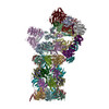
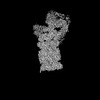
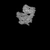
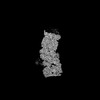
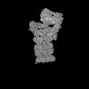
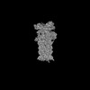
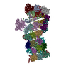
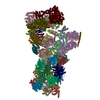
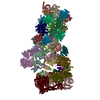
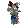
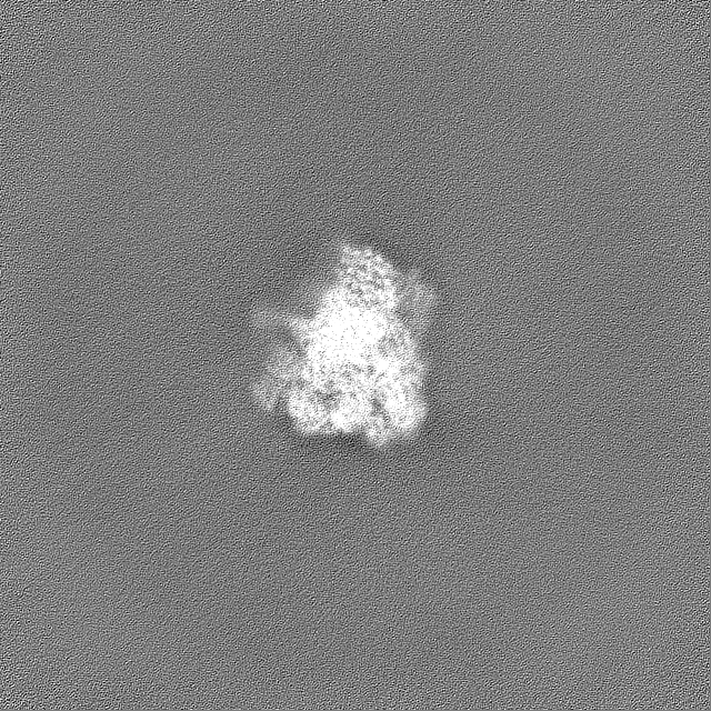
| Title | Locally Refined map of RP(19S) in substrate-engaged MIDN-bound 26S Proteasome, EB-MIDN state | |||||||||
 Map data Map data | ||||||||||
 Sample Sample |
| |||||||||
 Keywords Keywords | Ubiquitin-indepedent / proteasome / Midnolin / MIDN-bound 26S complex / IMMUNE SYSTEM | |||||||||
| Biological species |  Homo sapiens (human) Homo sapiens (human) | |||||||||
| Method | single particle reconstruction / cryo EM / Resolution: 2.8 Å | |||||||||
 Authors Authors | Peddada N / Beutler B | |||||||||
| Funding support |  United States, 2 items United States, 2 items
| |||||||||
 Citation Citation | Journal: Proc Natl Acad Sci U S A / Year: 2025 Title: Structural insights into the ubiquitin-independent midnolin-proteasome pathway. Authors: Nagesh Peddada / Xue Zhong / Yan Yin / Danielle Renee Lazaro / Jianhui Wang / Stephen Lyon / Jin Huk Choi / Xiao-Chen Bai / Eva Marie Y Moresco / Bruce Beutler / Abstract: The protein midnolin (MIDN) augments proteasome activity in lymphocytes and dramatically facilitates the survival and proliferation of B-lymphoid malignancies. MIDN binds both to proteasomes and to ...The protein midnolin (MIDN) augments proteasome activity in lymphocytes and dramatically facilitates the survival and proliferation of B-lymphoid malignancies. MIDN binds both to proteasomes and to substrates, but the mode of interaction with the proteasome is unknown, and the mechanism by which MIDN facilitates substrate degradation in a ubiquitin-independent manner is incompletely understood. Here, we present cryoelectron microscopy (cryo-EM) structures of the substrate-engaged, MIDN-bound human proteasome in two conformational states. MIDN induces proteasome conformations similarly to ubiquitinated substrates by using its ubiquitin-like domain to bind to the deubiquitinase RPN11 (PSMD14). By simultaneously binding to RPN1 (PSMD2) with its C-terminal α-helix, MIDN positions its substrate-carrying Catch domain above the proteasome ATPase channel through which substrates are translocated before degradation. Our findings suggest that both ubiquitin-like domain and C-terminal α-helix must bind to the proteasome for MIDN to stimulate proteasome activity. | |||||||||
| History |
|
- Structure visualization
| Supplemental images |
|---|

- Downloads & links
-EMDB archive
| Map data | emd_49498.map.gz | 883.3 MB |  EMDB map data format EMDB map data format | |
|---|---|---|---|---|
| Header (meta data) | emd-49498-v30.xmlemd-49498.xml | 21.4 KB 21.4 KB | Display Display | EMDB header |
| FSC (resolution estimation) | emd_49498_fsc.xml | 21 KB | Display | FSC data file |
| Images |  emd_49498.png emd_49498.png | 89.1 KB | ||
| Filedesc metadata | emd-49498.cif.gz | 4.8 KB | ||
| Others | emd_49498_half_map_1.map.gzemd_49498_half_map_2.map.gz | 929 MB 929 MB | ||
| Archive directory |  http://ftp.pdbj.org/pub/emdb/structures/EMD-49498ftp://ftp.pdbj.org/pub/emdb/structures/EMD-49498 http://ftp.pdbj.org/pub/emdb/structures/EMD-49498ftp://ftp.pdbj.org/pub/emdb/structures/EMD-49498 | HTTPS FTP |
-Related structure data
-Links
| EMDB pages | EMDB (EBI/PDBe) / EMDataResource |
|---|
-Map
| File | Download / File: emd_49498.map.gz / Format: CCP4 / Size: 1000 MB / Type: IMAGE STORED AS FLOATING POINT NUMBER (4 BYTES) | ||||||||||||||||||||||||||||||||||||
|---|---|---|---|---|---|---|---|---|---|---|---|---|---|---|---|---|---|---|---|---|---|---|---|---|---|---|---|---|---|---|---|---|---|---|---|---|---|
| Projections & slices | Image control
Images are generated by Spider. | ||||||||||||||||||||||||||||||||||||
| Voxel size | X=Y=Z: 1.074 Å | ||||||||||||||||||||||||||||||||||||
| Density |
| ||||||||||||||||||||||||||||||||||||
| Symmetry | Space group: 1 | ||||||||||||||||||||||||||||||||||||
| Details | EMDB XML:
|
 Z (Sec.)
 Y (Row.)
 X (Col.)


-Supplemental data
-Half map: #1
| File | emd_49498_half_map_1.map | ||||||||||||
|---|---|---|---|---|---|---|---|---|---|---|---|---|---|
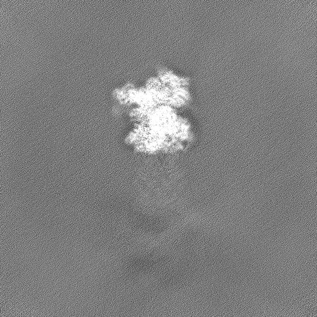
| Projections & Slices |
| ||||||||||||
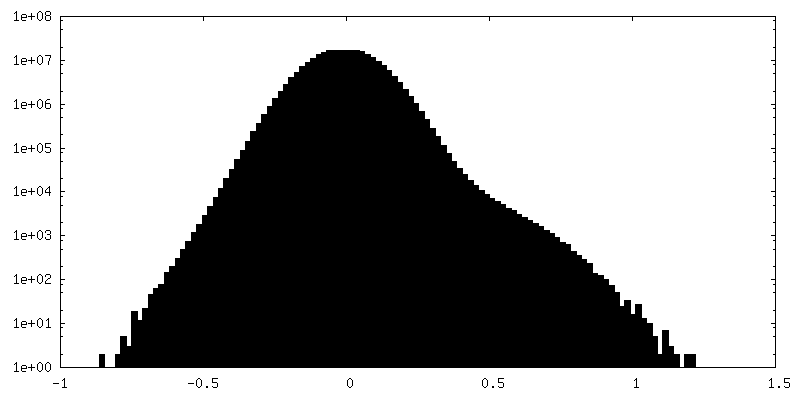
| Density Histograms |


-Half map: #2
| File | emd_49498_half_map_2.map | ||||||||||||
|---|---|---|---|---|---|---|---|---|---|---|---|---|---|
| Projections & Slices |
| ||||||||||||
| Density Histograms |


- Sample components
-Entire : Structure of MIDN-bound human 26S proteasome in substrate engaged...
| Entire | Name: Structure of MIDN-bound human 26S proteasome in substrate engaged state EB_MIDN |
|---|---|
| Components |
|
-Supramolecule #1: Structure of MIDN-bound human 26S proteasome in substrate engaged...
| Supramolecule | Name: Structure of MIDN-bound human 26S proteasome in substrate engaged state EB_MIDN type: complex / ID: 1 / Parent: 0 / Macromolecule list: #1-#32 |
|---|---|
| Source (natural) | Organism: Homo sapiens (human) / Strain: HEK 293 |
-Experimental details
-Structure determination
| Method | cryo EM |
|---|---|
 Processing Processing | single particle reconstruction |
| Aggregation state | particle |
-Sample preparation
| Buffer | pH: 7.6 Details: 50 mM Tris, pH 7.5, 150 mM NaCl, 20 mM KCl,5 mM MgCl2, 1 mM TECP, |
|---|---|
| Vitrification | Cryogen name: ETHANE / Chamber humidity: 100 % / Chamber temperature: 277 K / Instrument: FEI VITROBOT MARK IV |
| Details | In Vitro Reconstituted MIDN-26S proteasome complex |
- Electron microscopy
| Microscope | TFS KRIOS |
|---|---|
| Specialist optics | Energy filter - Name: GIF Bioquantum |
| Image recording | Film or detector model: GATAN K3 BIOQUANTUM (6k x 4k) / Number grids imaged: 3 / Number real images: 20794 / Average electron dose: 50.0 e/Å2 |
| Electron beam | Acceleration voltage: 300 kV / Electron source:  FIELD EMISSION GUN FIELD EMISSION GUN |
| Electron optics | C2 aperture diameter: 70.0 µm / Illumination mode: FLOOD BEAM / Imaging mode: BRIGHT FIELD / Cs: 2.7 mm / Nominal defocus max: 2.7 µm / Nominal defocus min: 1.1 µm / Nominal magnification: 81000 |
| Sample stage | Specimen holder model: FEI TITAN KRIOS AUTOGRID HOLDER |
| Experimental equipment |  Model: Titan Krios / Image courtesy: FEI Company |
+Image processing

-Atomic model buiding 1
| Initial model | PDB ID: Chain - Source name: PDB / Chain - Initial model type: experimental model |
|---|---|
| Refinement | Space: REAL / Protocol: RIGID BODY FIT |