National Natural Science Foundation of China (NSFC)
12034006
China
National Natural Science Foundation of China (NSFC)
32071209
China
National Natural Science Foundation of China (NSFC)
32200994
China
Citation
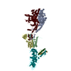
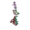
Journal: Structure / Year: 2024 Title: Three-dimensional structures of Vibrio cholerae typing podophage VP1 in two states. Authors: Hao Pang / Fenxia Fan / Jing Zheng / Hao Xiao / Zhixue Tan / Jingdong Song / Biao Kan / Hongrong Liu / Abstract: Lytic podophages (VP1-VP5) play crucial roles in subtyping Vibrio cholerae O1 biotype El Tor. However, until now no structures of these phages have been available, which hindered our understanding of ...Lytic podophages (VP1-VP5) play crucial roles in subtyping Vibrio cholerae O1 biotype El Tor. However, until now no structures of these phages have been available, which hindered our understanding of the molecular mechanisms of infection and DNA release. Here, we determined the cryoelectron microscopy (cryo-EM) structures of mature and DNA-ejected VP1 structures at near-atomic and subnanometer resolutions, respectively. The VP1 head is composed of 415 copies of the major capsid protein gp7 and 11 turret-shaped spikes. The VP1 tail consists of an adapter, a nozzle, a slender ring, and a tail needle, and is flanked by three extended fibers I and six trimeric fibers II. Conformational changes of fiber II in DNA-ejected VP1 may cause the release of the tail needle and core proteins, forming an elongated tail channel. Our structures provide insights into the molecular mechanisms of infection and DNA release for podophages with a tail needle.
History
Deposition
Jul 5, 2024
Deposition site: PDBJ / Processing site: PDBJ
Revision 1.0
Aug 14, 2024
Provider: repository / Type: Initial release
Revision 1.0
Aug 14, 2024
Data content type: EM metadata / Data content type: EM metadata / Provider: repository / Type: Initial release
Revision 1.0
Aug 14, 2024
Data content type: Half map / Part number: 1 / Data content type: Half map / Provider: repository / Type: Initial release
Revision 1.0
Aug 14, 2024
Data content type: Half map / Part number: 2 / Data content type: Half map / Provider: repository / Type: Initial release
Revision 1.0
Aug 14, 2024
Data content type: Image / Data content type: Image / Provider: repository / Type: Initial release
Revision 1.0
Aug 14, 2024
Data content type: Primary map / Data content type: Primary map / Provider: repository / Type: Initial release
Revision 1.0
Aug 14, 2024
Data content type: Half map / Part number: 1 / Data content type: Half map / Provider: repository / Type: Initial release
Revision 1.0
Aug 14, 2024
Data content type: Half map / Part number: 2 / Data content type: Half map / Provider: repository / Type: Initial release
Revision 1.0
Aug 14, 2024
Data content type: Image / Data content type: Image / Provider: repository / Type: Initial release
Revision 1.0
Aug 14, 2024
Data content type: Primary map / Data content type: Primary map / Provider: repository / Type: Initial release
Data content type: EM metadata / Data content type: EM metadata / EM metadata / Group: Data processing / Experimental summary / Data content type: EM metadata / EM metadata / Category: em_admin / em_software / Data content type: EM metadata / EM metadata / Item: _em_admin.last_update / _em_software.name
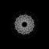
M: major capsid of VP1 N: major capsid of VP1 O: major capsid of VP1 P: major capsid of VP1 Q: major capsid of VP1 R: major capsid of VP1 T: major capsid of VP1 c: SHP of VP1
In the structure databanks used in Yorodumi, some data are registered as the other names, "COVID-19 virus" and "2019-nCoV". Here are the details of the virus and the list of structure data.
Jan 31, 2019. EMDB accession codes are about to change! (news from PDBe EMDB page)
EMDB accession codes are about to change! (news from PDBe EMDB page)
The allocation of 4 digits for EMDB accession codes will soon come to an end. Whilst these codes will remain in use, new EMDB accession codes will include an additional digit and will expand incrementally as the available range of codes is exhausted. The current 4-digit format prefixed with “EMD-” (i.e. EMD-XXXX) will advance to a 5-digit format (i.e. EMD-XXXXX), and so on. It is currently estimated that the 4-digit codes will be depleted around Spring 2019, at which point the 5-digit format will come into force.
The EM Navigator/Yorodumi systems omit the EMD- prefix.
Related info.:Q: What is EMD? / ID/Accession-code notation in Yorodumi/EM Navigator
Yorodumi is a browser for structure data from EMDB, PDB, SASBDB, etc.
This page is also the successor to EM Navigator detail page, and also detail information page/front-end page for Omokage search.
The word "yorodu" (or yorozu) is an old Japanese word meaning "ten thousand". "mi" (miru) is to see.
Related info.:EMDB / PDB / SASBDB / Comparison of 3 databanks / Yorodumi Search / Aug 31, 2016. New EM Navigator & Yorodumi / Yorodumi Papers / Jmol/JSmol / Function and homology information / Changes in new EM Navigator and Yorodumi
 Movie
Movie Controller


 Open data
 Basic information
 Components
 Keywords
 Vibrio cholerae (bacteria)
 Authors
 China, 3items
  Citation
 Structure visualization
 Molmil
 Downloads & links
 Other downloads

 PDBj
PDBj Assembly
 Sample preparation
 Electron microscopy imaging
 FIELD EMISSION GUN / Accelerating voltage: 300 kV / Illumination mode: FLOOD BEAM
 Processing