 Movie
Movie Controller


+ Open data

- Basic information
| Entry | Database: PDB / ID: 8zkk | ||||||||||||
|---|---|---|---|---|---|---|---|---|---|---|---|---|---|
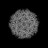
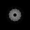
| Title | Portal-tail of Vibrio cholerae typing phage mature VP1 | ||||||||||||
 Components Components |
| ||||||||||||
 Keywords Keywords | VIRAL PROTEIN / phage / virus / vibrio cholera phage | ||||||||||||
| Biological species |   Vibrio cholerae (bacteria) Vibrio cholerae (bacteria) | ||||||||||||
| Method | ELECTRON MICROSCOPY / single particle reconstruction / cryo EM / Resolution: 3.6 Å | ||||||||||||
 Authors Authors | Liu, H.R. / Pang, H. | ||||||||||||
| Funding support |  China, 3items China, 3items
| ||||||||||||
 Citation Citation | Journal: Structure / Year: 2024 Title: Three-dimensional structures of Vibrio cholerae typing podophage VP1 in two states. Authors: Hao Pang / Fenxia Fan / Jing Zheng / Hao Xiao / Zhixue Tan / Jingdong Song / Biao Kan / Hongrong Liu / Abstract: Lytic podophages (VP1-VP5) play crucial roles in subtyping Vibrio cholerae O1 biotype El Tor. However, until now no structures of these phages have been available, which hindered our understanding of ...Lytic podophages (VP1-VP5) play crucial roles in subtyping Vibrio cholerae O1 biotype El Tor. However, until now no structures of these phages have been available, which hindered our understanding of the molecular mechanisms of infection and DNA release. Here, we determined the cryoelectron microscopy (cryo-EM) structures of mature and DNA-ejected VP1 structures at near-atomic and subnanometer resolutions, respectively. The VP1 head is composed of 415 copies of the major capsid protein gp7 and 11 turret-shaped spikes. The VP1 tail consists of an adapter, a nozzle, a slender ring, and a tail needle, and is flanked by three extended fibers I and six trimeric fibers II. Conformational changes of fiber II in DNA-ejected VP1 may cause the release of the tail needle and core proteins, forming an elongated tail channel. Our structures provide insights into the molecular mechanisms of infection and DNA release for podophages with a tail needle. | ||||||||||||
| History |
|
- Structure visualization
| Structure viewer | Molecule:  MolmilJmol/JSmol MolmilJmol/JSmol |
|---|
- Downloads & links
-Download
| PDBx/mmCIF format | 8zkk.cif.gz | 409.9 KB | Display | PDBx/mmCIF format |
|---|---|---|---|---|
| PDB format | pdb8zkk.ent.gz | 332.2 KB | Display | PDB format |
| PDBx/mmJSON format | 8zkk.json.gz | Tree view | PDBx/mmJSON format | |
| Others |  Other downloads Other downloads |
-Validation report
| Arichive directory | https://data.pdbj.org/pub/pdb/validation_reports/zk/8zkkftp://data.pdbj.org/pub/pdb/validation_reports/zk/8zkk | HTTPS FTP |
|---|
-Related structure data
| Related structure data | 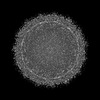 60197MC 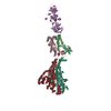 8zkmC 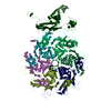 9in6C M: map data used to model this data C: citing same article ( |
|---|


-Links
 PDBj
PDBj- Assembly
| Deposited unit | 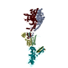
|
|---|---|
| 1 | x 6
|
-Components
| #1: Protein | Mass: 59015.762 Da / Num. of mol.: 1 / Source method: isolated from a natural source / Source: (natural) Vibrio cholerae (bacteria) | ||||||||
|---|---|---|---|---|---|---|---|---|---|
| #2: Protein | Mass: 73263.602 Da / Num. of mol.: 2 / Source method: isolated from a natural source / Source: (natural) Vibrio cholerae (bacteria)#3: Protein | Mass: 23592.850 Da / Num. of mol.: 2 / Source method: isolated from a natural source / Source: (natural) Vibrio cholerae (bacteria)#4: Protein | | Mass: 26311.119 Da / Num. of mol.: 1 / Source method: isolated from a natural source / Source: (natural) Vibrio cholerae (bacteria)#5: Protein | Mass: 10025.203 Da / Num. of mol.: 3 / Source method: isolated from a natural source / Source: (natural) Vibrio cholerae (bacteria)Has protein modification | N | |
-Experimental details
-Experiment
| Experiment | Method: ELECTRON MICROSCOPY |
|---|---|
| EM experiment | Aggregation state: PARTICLE / 3D reconstruction method: single particle reconstruction |
- Sample preparation
| Component | Name: Vibrio cholerae phage / Type: VIRUS / Entity ID: all / Source: NATURAL |
|---|---|
| Source (natural) | Organism: Vibrio cholerae phage (bacteria) |
| Details of virus | Empty: NO / Enveloped: NO / Isolate: SPECIES / Type: VIRION |
| Buffer solution | pH: 7.4 |
| Specimen | Embedding applied: NO / Shadowing applied: NO / Staining applied: NO / Vitrification applied: YES |
| Vitrification | Cryogen name: ETHANE |
- Electron microscopy imaging
| Experimental equipment |  Model: Titan Krios / Image courtesy: FEI Company |
|---|---|
| Microscopy | Model: FEI TITAN KRIOS |
| Electron gun | Electron source:  FIELD EMISSION GUN / Accelerating voltage: 300 kV / Illumination mode: FLOOD BEAM FIELD EMISSION GUN / Accelerating voltage: 300 kV / Illumination mode: FLOOD BEAM |
| Electron lens | Mode: BRIGHT FIELD / Nominal defocus max: 2200 nm / Nominal defocus min: 1600 nm |
| Image recording | Electron dose: 32 e/Å2 / Film or detector model: GATAN K3 (6k x 4k) |
- Processing
| CTF correction | Type: PHASE FLIPPING AND AMPLITUDE CORRECTION |
|---|---|
| 3D reconstruction | Resolution: 3.6 Å / Resolution method: FSC 0.143 CUT-OFF / Num. of particles: 47456 / Symmetry type: POINT |
