 Movie
Movie Controller

[English] 日本語
 Yorodumi
- PDB-9e0q: CryoEM structure of inducible Lysine decarboxylase from Hafnia al... -
+ Open data

- Basic information
| Entry | Database: PDB / ID: 9e0q | |||||||||||||||
|---|---|---|---|---|---|---|---|---|---|---|---|---|---|---|---|---|
| Title | CryoEM structure of inducible Lysine decarboxylase from Hafnia alvei D-hydrazino-Lysine analog at 2.3 Angstrom resolution | |||||||||||||||
 Components Components | Lysine decarboxylase, inducible | |||||||||||||||
 Keywords Keywords | LYASE / L-Lysine decarboxylase / cadaverine / D-alpha-hydrazone inhibitor | |||||||||||||||
| Function / homology |  Function and homology information Function and homology informationarginine decarboxylase activity / lysine decarboxylase / lysine decarboxylase activity / : / L-arginine catabolic process / pyridoxal phosphate binding / identical protein binding / cytosol Similarity search - Function | |||||||||||||||
| Biological species |  Hafnia alvei ATCC 51873 (bacteria) Hafnia alvei ATCC 51873 (bacteria) | |||||||||||||||
| Method | ELECTRON MICROSCOPY / single particle reconstruction / cryo EM / Resolution: 2.3 Å | |||||||||||||||
 Authors Authors | Duhoo, Y. / Desfosses, A. / Gutsche, I. / Doukov, T.I. / Berkowitz, D.B. | |||||||||||||||
| Funding support |  France, France,  United States, 4items United States, 4items
| |||||||||||||||
 Citation Citation | Journal: ACS Catal / Year: 2025 Title: α-Hydrazino Acids Inhibit Pyridoxal Phosphate-Dependent Decarboxylases via "Catalytically Correct" Ketoenamine Tautomers: A Special Motif for Chemical Biology and Drug Discovery? Authors: Jonathan M Baine / Yoan Duhoo / Tzanko Doukov / Ambroise Desfosses / Giovanni Bisello / Matthew L Beio / Olivia Bauer / Massimiliano Perduca / Maria Bacia-Verloop / Mariarita Bertoldi / ...Authors: Jonathan M Baine / Yoan Duhoo / Tzanko Doukov / Ambroise Desfosses / Giovanni Bisello / Matthew L Beio / Olivia Bauer / Massimiliano Perduca / Maria Bacia-Verloop / Mariarita Bertoldi / Robert S Phillips / Irina Gutsche / David B Berkowitz /   Abstract: We present evidence that supports a 'correct hydrazone tautomer/Dunathan alignment model' for how α-hydrazino analogues of α-amino acids inhibit PLP enzymes. Described is the asymmetric synthesis ...We present evidence that supports a 'correct hydrazone tautomer/Dunathan alignment model' for how α-hydrazino analogues of α-amino acids inhibit PLP enzymes. Described is the asymmetric synthesis of l- and d-α-hydrazino acid l-lysine analogues and their inhibition of lysine decarboxylase (LdcI) via kinetic analysis, stopped-flow spectrophotometry, and cryo-EM. We describe a similar investigation of the important anti-Parkinsonism drug, carbidopa, with its human DOPA decarboxylase (hDdc) target. Evidence is consistent with these three hydrazino analogues forming the catalytically relevant ketoenamine PLP-hydrazone tautomer in their target active sites, with the α-carboxylate groups, though insulated, aligning with the PLP-π-system in a Dunathan-model-like orientation. High-resolution cryo-EM structures of the LdcI holoenzyme (pdb 9E0M-2.1Å) and LdcI-bound l- and d-hydrazones (pdb 9E0O-2.0 Å; pdb 9E0Q-2.3Å) and the first X-ray crystal structure of hDdc-bound carbidopa (pdb 9GNS-1.93Å) support this 'correct tautomer' model. These insights are expected to guide future PLP enzyme inhibitor development. #1: Journal: Acta Crystallogr D Struct Biol / Year: 2019 Title: Macromolecular structure determination using X-rays, neutrons and electrons: recent developments in Phenix. Authors: Dorothee Liebschner / Pavel V Afonine / Matthew L Baker / Gábor Bunkóczi / Vincent B Chen / Tristan I Croll / Bradley Hintze / Li Wei Hung / Swati Jain / Airlie J McCoy / Nigel W Moriarty ...Authors: Dorothee Liebschner / Pavel V Afonine / Matthew L Baker / Gábor Bunkóczi / Vincent B Chen / Tristan I Croll / Bradley Hintze / Li Wei Hung / Swati Jain / Airlie J McCoy / Nigel W Moriarty / Robert D Oeffner / Billy K Poon / Michael G Prisant / Randy J Read / Jane S Richardson / David C Richardson / Massimo D Sammito / Oleg V Sobolev / Duncan H Stockwell / Thomas C Terwilliger / Alexandre G Urzhumtsev / Lizbeth L Videau / Christopher J Williams / Paul D Adams /  Abstract: Diffraction (X-ray, neutron and electron) and electron cryo-microscopy are powerful methods to determine three-dimensional macromolecular structures, which are required to understand biological ...Diffraction (X-ray, neutron and electron) and electron cryo-microscopy are powerful methods to determine three-dimensional macromolecular structures, which are required to understand biological processes and to develop new therapeutics against diseases. The overall structure-solution workflow is similar for these techniques, but nuances exist because the properties of the reduced experimental data are different. Software tools for structure determination should therefore be tailored for each method. Phenix is a comprehensive software package for macromolecular structure determination that handles data from any of these techniques. Tasks performed with Phenix include data-quality assessment, map improvement, model building, the validation/rebuilding/refinement cycle and deposition. Each tool caters to the type of experimental data. The design of Phenix emphasizes the automation of procedures, where possible, to minimize repetitive and time-consuming manual tasks, while default parameters are chosen to encourage best practice. A graphical user interface provides access to many command-line features of Phenix and streamlines the transition between programs, project tracking and re-running of previous tasks. | |||||||||||||||
| History |
|
- Structure visualization
| Structure viewer | Molecule: MolmilJmol/JSmol |
|---|
- Downloads & links
-Download
| PDBx/mmCIF format | 9e0q.cif.gz | 1.6 MB | Display | PDBx/mmCIF format |
|---|---|---|---|---|
| PDB format | pdb9e0q.ent.gz | Display | PDB format | |
| PDBx/mmJSON format | 9e0q.json.gz | Tree view | PDBx/mmJSON format | |
| Others |  Other downloads Other downloads |
-Validation report
| Arichive directory | https://data.pdbj.org/pub/pdb/validation_reports/e0/9e0qftp://data.pdbj.org/pub/pdb/validation_reports/e0/9e0q | HTTPS FTP |
|---|
-Related structure data
| Related structure data |  47366MC  9duiC  9e0mC  9e0oC  9gnsC M: map data used to model this data C: citing same article ( |
|---|---|
| Similar structure data |


-Links
 PDBj
PDBj- Assembly
| Deposited unit | 
|
|---|---|
| 1 |
|
-Components
| #1: Protein | Mass: 80205.281 Da / Num. of mol.: 10 Source method: isolated from a genetically manipulated source Source: (gene. exp.) Hafnia alvei ATCC 51873 (bacteria) / Gene: HMPREF0454_03276Production host: References: UniProt: G9Y9L1 #2: Chemical | ChemComp-A1BD0 / ( Mass: 390.329 Da / Num. of mol.: 10 / Source method: obtained synthetically / Formula: C14H23N4O7P / Feature type: SUBJECT OF INVESTIGATION #3: Water | ChemComp-HOH / |  Mass: 18.015 Da / Num. of mol.: 727 / Source method: isolated from a natural source / Formula: H2O Mass: 18.015 Da / Num. of mol.: 727 / Source method: isolated from a natural source / Formula: H2OHas ligand of interest | Y | Has protein modification | N | |
|---|
-Experimental details
-Experiment
| Experiment | Method: ELECTRON MICROSCOPY |
|---|---|
| EM experiment | Aggregation state: PARTICLE / 3D reconstruction method: single particle reconstruction |
- Sample preparation
| Component | Name: Inducible Lysine decarboxylase from Hafnia alvei with D-alpha-hydrazino-Lysine inhibitor Type: COMPLEX Details: Inducible Lysine decarboxylase from Hafnia alvei with D-alpha-hydrazino-Lysine inhibitor in ketoenamine form Entity ID: #1 / Source: RECOMBINANT |
|---|---|
| Source (natural) | Organism: Hafnia alvei ATCC 51873 (bacteria) |
| Source (recombinant) | Organism: Plasmid: peT28+ |
| Buffer solution | pH: 7.4 |
| Specimen | Conc.: 1 mg/ml / Embedding applied: NO / Shadowing applied: NO / Staining applied: NO / Vitrification applied: YES |
| Specimen support | Grid material: COPPER / Grid mesh size: 300 divisions/in. / Grid type: Quantifoil R2/1 |
| Vitrification | Instrument: FEI VITROBOT MARK IV / Cryogen name: ETHANE / Humidity: 100 % / Chamber temperature: 295 K |
- Electron microscopy imaging
| Microscopy | Model: TFS GLACIOS |
|---|---|
| Electron gun | Electron source:  FIELD EMISSION GUN / Accelerating voltage: 200 kV / Illumination mode: FLOOD BEAM FIELD EMISSION GUN / Accelerating voltage: 200 kV / Illumination mode: FLOOD BEAM |
| Electron lens | Mode: BRIGHT FIELD / Nominal defocus max: 2200 nm / Nominal defocus min: 800 nm |
| Image recording | Electron dose: 40 e/Å2 / Film or detector model: GATAN K2 SUMMIT (4k x 4k) |
- Processing
| EM software |
| ||||||||||||||||||||||||
|---|---|---|---|---|---|---|---|---|---|---|---|---|---|---|---|---|---|---|---|---|---|---|---|---|---|
| CTF correction | Type: PHASE FLIPPING AND AMPLITUDE CORRECTION | ||||||||||||||||||||||||
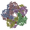
| Symmetry | Point symmetry: D5 (2x5 fold dihedral) | ||||||||||||||||||||||||
| 3D reconstruction | Resolution: 2.3 Å / Resolution method: FSC 0.143 CUT-OFF / Num. of particles: 360000 / Symmetry type: POINT | ||||||||||||||||||||||||
| Refinement | Cross valid method: NONE Stereochemistry target values: GeoStd + Monomer Library + CDL v1.2 | ||||||||||||||||||||||||
| Displacement parameters | Biso mean: 13.23 Å2 | ||||||||||||||||||||||||
| Refine LS restraints |
|
