 Movie
Movie Controller

[English] 日本語
 Yorodumi
- PDB-9dui: Re-refined of Crystal structure of dopa decarboxylase in complex ... -
+ Open data

- Basic information
| Entry | Database: PDB / ID: 9dui | |||||||||
|---|---|---|---|---|---|---|---|---|---|---|
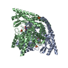
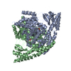
| Title | Re-refined of Crystal structure of dopa decarboxylase in complex with the inhibitor carbidopa (1JS3) with ketoenamine form of carbidopa | |||||||||
 Components Components | Aromatic-L-amino-acid decarboxylase | |||||||||
 Keywords Keywords | LYASE / DOPA decarboxylase / carbiDOPA / Parkinson's disease / Vitamin B6 | |||||||||
| Function / homology |  Function and homology information Function and homology informationL-dopa decarboxylase activity / 5-hydroxy-L-tryptophan decarboxylase activity / aromatic-L-amino-acid decarboxylase / aromatic-L-amino-acid decarboxylase activity / serotonin biosynthetic process / catecholamine metabolic process / carboxylic acid metabolic process / amino acid metabolic process / dopamine biosynthetic process / pyridoxal phosphate binding / cytoplasm Similarity search - Function | |||||||||
| Biological species |  | |||||||||
| Method |  X-RAY DIFFRACTION / SYNCHROTRON / MAD / Resolution: 2.25 Å X-RAY DIFFRACTION / SYNCHROTRON / MAD / Resolution: 2.25 Å | |||||||||
 Authors Authors | Doukov, I.T. / Berkowitz, B.D. | |||||||||
| Funding support |  United States, 2items United States, 2items
| |||||||||
 Citation Citation | Journal: Nat.Struct.Biol. / Year: 2001 Title: Structural insight into Parkinson's disease treatment from drug-inhibited DOPA decarboxylase. Authors: Burkhard, P. / Dominici, P. / Borri-Voltattorni, C. / Jansonius, J.N. / Malashkevich, V.N. #1: Journal: Acta Crystallogr.,Sect.D / Year: 1999 Title: Preliminary X-ray analysis of a new crystal form of pig kidney DOPA decarboxylase Authors: Malashkevich, V.N. / Burkhard, P. / Dominici, P. / Moore, P.S. / Borri-Voltattorni, C. / Jansonius, J.N. #2: Journal: J.Mol.Biol. / Year: 1992 Title: Crystallization and preliminary X-ray analysis of pig kidney DOPA decarboxylase Authors: Malashkevich, V.N. / Filipponi, P. / Sauder, U. / Dominici, P. / Jansonius, J.N. / Borri-Voltattorni, C. | |||||||||
| History |
| |||||||||
| Remark 0 | THIS ENTRY 9DUI REFLECTS AN ALTERNATIVE MODELING OF THE ORIGINAL DATA IN 1JS3, DETERMINED BY P. ...THIS ENTRY 9DUI REFLECTS AN ALTERNATIVE MODELING OF THE ORIGINAL DATA IN 1JS3, DETERMINED BY P.BURKHARD,C.BORRI-VOLTATTORNI,J.N.JANSONIUS, V.N.MALASHKEVICH |
- Structure visualization
| Structure viewer | Molecule: MolmilJmol/JSmol |
|---|
- Downloads & links
-Download
| PDBx/mmCIF format | 9dui.cif.gz | 501.2 KB | Display | PDBx/mmCIF format |
|---|---|---|---|---|
| PDB format | pdb9dui.ent.gz | Display | PDB format | |
| PDBx/mmJSON format | 9dui.json.gz | Tree view | PDBx/mmJSON format | |
| Others |  Other downloads Other downloads |
-Validation report
| Arichive directory | https://data.pdbj.org/pub/pdb/validation_reports/du/9duiftp://data.pdbj.org/pub/pdb/validation_reports/du/9dui | HTTPS FTP |
|---|
-Related structure data


-Links
 PDBj
PDBj- Assembly
| Deposited unit | 
| ||||||||||||
|---|---|---|---|---|---|---|---|---|---|---|---|---|---|
| 1 |
| ||||||||||||
| Unit cell |
| ||||||||||||
| Noncrystallographic symmetry (NCS) | NCS domain:
NCS domain segments: Component-ID: 1 / Ens-ID: 1 / Beg auth comp-ID: ASN / Beg label comp-ID: ASN / End auth comp-ID: GLU / End label comp-ID: GLU / Auth asym-ID: A / Label asym-ID: A / Auth seq-ID: 2 - 478 / Label seq-ID: 3 - 479
NCS ensembles : (Details: Local NCS retraints between domains: 1 2) |
-Components
| #1: Protein | Mass: 54028.211 Da / Num. of mol.: 2 Source method: isolated from a genetically manipulated source Source: (gene. exp.)  References: UniProt: P80041, aromatic-L-amino-acid decarboxylase #2: Chemical | Mass: 455.356 Da / Num. of mol.: 2 / Source method: obtained synthetically / Formula: C18H22N3O9P / Feature type: SUBJECT OF INVESTIGATION #3: Chemical | ChemComp-SO4 /   Mass: 96.063 Da / Num. of mol.: 4 / Source method: obtained synthetically / Formula: SO4 Mass: 96.063 Da / Num. of mol.: 4 / Source method: obtained synthetically / Formula: SO4#4: Water | ChemComp-HOH / |  Mass: 18.015 Da / Num. of mol.: 579 / Source method: isolated from a natural source / Formula: H2O Mass: 18.015 Da / Num. of mol.: 579 / Source method: isolated from a natural source / Formula: H2OHas ligand of interest | Y | Has protein modification | Y | |
|---|
-Experimental details
-Experiment
| Experiment | Method: X-RAY DIFFRACTION / Number of used crystals: 1 |
|---|
- Sample preparation
| Crystal | Density Matthews: 2.76 Å3/Da / Density % sol: 55.47 % / Description: AUTHOR USED THE SF DATA FROM ENTRY 1J3S. |
|---|---|
| Crystal grow | Temperature: 298 K / Method: vapor diffusion, hanging drop / pH: 6.5 Details: PEG MME 5000, MES, ammonium sulfate at pH 6.5, VAPOR DIFFUSION, HANGING DROP at 298K, temperature 298.0K |
-Data collection
| Diffraction | Mean temperature: 100 K / Serial crystal experiment: N |
|---|---|
| Diffraction source | Source: SYNCHROTRON / Site: LURE  / Beamline: D41A / Wavelength: 0.95 Å / Beamline: D41A / Wavelength: 0.95 Å |
| Detector | Type: MARRESEARCH / Detector: IMAGE PLATE / Date: Apr 10, 1993 |
| Radiation | Protocol: SINGLE WAVELENGTH / Monochromatic (M) / Laue (L): M / Scattering type: x-ray |
| Radiation wavelength | Wavelength: 0.95 Å / Relative weight: 1 |
| Reflection | Resolution: 2.25→19.611 Å / Num. obs: 53582 / % possible obs: 95.8 % / Observed criterion σ(I): 0 / Redundancy: 2.9 % / Biso Wilson estimate: 15 Å2 / Rsym value: 0.056 / Net I/σ(I): 9.5 |
| Reflection shell | Resolution: 2.25→2.3 Å / Redundancy: 1.7 % / Mean I/σ(I) obs: 4.7 / Num. unique obs: 154 / Rsym value: 0.19 / % possible all: 70.7 |
- Processing
| Software |
| |||||||||||||||||||||||||||||||||||||||||||||||||||||||||||||||||||||||||||||||||||||||||||||||||||||||||||||||||||||||||||||||||||||||||||||||||||||||||||
|---|---|---|---|---|---|---|---|---|---|---|---|---|---|---|---|---|---|---|---|---|---|---|---|---|---|---|---|---|---|---|---|---|---|---|---|---|---|---|---|---|---|---|---|---|---|---|---|---|---|---|---|---|---|---|---|---|---|---|---|---|---|---|---|---|---|---|---|---|---|---|---|---|---|---|---|---|---|---|---|---|---|---|---|---|---|---|---|---|---|---|---|---|---|---|---|---|---|---|---|---|---|---|---|---|---|---|---|---|---|---|---|---|---|---|---|---|---|---|---|---|---|---|---|---|---|---|---|---|---|---|---|---|---|---|---|---|---|---|---|---|---|---|---|---|---|---|---|---|---|---|---|---|---|---|---|---|
| Refinement | Method to determine structure: MAD / Resolution: 2.25→19.61 Å / Cor.coef. Fo:Fc: 0.977 / Cor.coef. Fo:Fc free: 0.958 / SU B: 7.863 / SU ML: 0.096 / Cross valid method: THROUGHOUT / ESU R: 0.176 / ESU R Free: 0.141 Details: Hydrogens have been added in their riding positions
| |||||||||||||||||||||||||||||||||||||||||||||||||||||||||||||||||||||||||||||||||||||||||||||||||||||||||||||||||||||||||||||||||||||||||||||||||||||||||||
| Solvent computation | Ion probe radii: 0.8 Å / Shrinkage radii: 0.8 Å / VDW probe radii: 1.2 Å / Solvent model: MASK BULK SOLVENT | |||||||||||||||||||||||||||||||||||||||||||||||||||||||||||||||||||||||||||||||||||||||||||||||||||||||||||||||||||||||||||||||||||||||||||||||||||||||||||
| Displacement parameters | Biso mean: 23.854 Å2
| |||||||||||||||||||||||||||||||||||||||||||||||||||||||||||||||||||||||||||||||||||||||||||||||||||||||||||||||||||||||||||||||||||||||||||||||||||||||||||
| Refinement step | Cycle: LAST / Resolution: 2.25→19.61 Å
| |||||||||||||||||||||||||||||||||||||||||||||||||||||||||||||||||||||||||||||||||||||||||||||||||||||||||||||||||||||||||||||||||||||||||||||||||||||||||||
| Refine LS restraints |
| |||||||||||||||||||||||||||||||||||||||||||||||||||||||||||||||||||||||||||||||||||||||||||||||||||||||||||||||||||||||||||||||||||||||||||||||||||||||||||
| Refine LS restraints NCS |
| |||||||||||||||||||||||||||||||||||||||||||||||||||||||||||||||||||||||||||||||||||||||||||||||||||||||||||||||||||||||||||||||||||||||||||||||||||||||||||
| LS refinement shell |
| |||||||||||||||||||||||||||||||||||||||||||||||||||||||||||||||||||||||||||||||||||||||||||||||||||||||||||||||||||||||||||||||||||||||||||||||||||||||||||
| Refinement TLS params. | Method: refined / Refine-ID: X-RAY DIFFRACTION
| |||||||||||||||||||||||||||||||||||||||||||||||||||||||||||||||||||||||||||||||||||||||||||||||||||||||||||||||||||||||||||||||||||||||||||||||||||||||||||
| Refinement TLS group | Selection: ALL |
