 Movie
Movie Controller

[English] 日本語
 Yorodumi
- PDB-9e0m: CryoEM structure of holoenzyme of inducible Lysine decarboxylase ... -
+ Open data

- Basic information
| Entry | Database: PDB / ID: 9e0m | |||||||||||||||
|---|---|---|---|---|---|---|---|---|---|---|---|---|---|---|---|---|
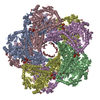
| Title | CryoEM structure of holoenzyme of inducible Lysine decarboxylase from Hafnia alvei holoenzyme at 2.19 Angstrom resolution | |||||||||||||||
 Components Components | Lysine decarboxylase, inducible | |||||||||||||||
 Keywords Keywords | LYASE / L-lysine carboxylyase / pH homeostasis / cadaverine / pyridoxal-phosphate protein | |||||||||||||||
| Function / homology |  Function and homology information Function and homology informationarginine decarboxylase activity / lysine decarboxylase / lysine decarboxylase activity / : / L-arginine catabolic process / pyridoxal phosphate binding / identical protein binding / cytosol Similarity search - Function | |||||||||||||||
| Biological species |  Hafnia alvei ATCC 51873 (bacteria) Hafnia alvei ATCC 51873 (bacteria) | |||||||||||||||
| Method | ELECTRON MICROSCOPY / single particle reconstruction / cryo EM / Resolution: 2.2 Å | |||||||||||||||
 Authors Authors | Duhoo, Y. / Desfosses, A. / Gutsche, I. / Doukov, T.I. / Berkowitz, D.B. | |||||||||||||||
| Funding support |  France, France,  United States, 4items United States, 4items
| |||||||||||||||
 Citation Citation | Journal: ACS Catal / Year: 2025 Title: α-Hydrazino Acids Inhibit Pyridoxal Phosphate-Dependent Decarboxylases via "Catalytically Correct" Ketoenamine Tautomers: A Special Motif for Chemical Biology and Drug Discovery? Authors: Jonathan M Baine / Yoan Duhoo / Tzanko Doukov / Ambroise Desfosses / Giovanni Bisello / Matthew L Beio / Olivia Bauer / Massimiliano Perduca / Maria Bacia-Verloop / Mariarita Bertoldi / ...Authors: Jonathan M Baine / Yoan Duhoo / Tzanko Doukov / Ambroise Desfosses / Giovanni Bisello / Matthew L Beio / Olivia Bauer / Massimiliano Perduca / Maria Bacia-Verloop / Mariarita Bertoldi / Robert S Phillips / Irina Gutsche / David B Berkowitz /   Abstract: We present evidence that supports a 'correct hydrazone tautomer/Dunathan alignment model' for how α-hydrazino analogues of α-amino acids inhibit PLP enzymes. Described is the asymmetric synthesis ...We present evidence that supports a 'correct hydrazone tautomer/Dunathan alignment model' for how α-hydrazino analogues of α-amino acids inhibit PLP enzymes. Described is the asymmetric synthesis of l- and d-α-hydrazino acid l-lysine analogues and their inhibition of lysine decarboxylase (LdcI) via kinetic analysis, stopped-flow spectrophotometry, and cryo-EM. We describe a similar investigation of the important anti-Parkinsonism drug, carbidopa, with its human DOPA decarboxylase (hDdc) target. Evidence is consistent with these three hydrazino analogues forming the catalytically relevant ketoenamine PLP-hydrazone tautomer in their target active sites, with the α-carboxylate groups, though insulated, aligning with the PLP-π-system in a Dunathan-model-like orientation. High-resolution cryo-EM structures of the LdcI holoenzyme (pdb 9E0M-2.1Å) and LdcI-bound l- and d-hydrazones (pdb 9E0O-2.0 Å; pdb 9E0Q-2.3Å) and the first X-ray crystal structure of hDdc-bound carbidopa (pdb 9GNS-1.93Å) support this 'correct tautomer' model. These insights are expected to guide future PLP enzyme inhibitor development. | |||||||||||||||
| History |
|
- Structure visualization
| Structure viewer | Molecule: MolmilJmol/JSmol |
|---|
- Downloads & links
-Download
| PDBx/mmCIF format | 9e0m.cif.gz | 1.7 MB | Display | PDBx/mmCIF format |
|---|---|---|---|---|
| PDB format | pdb9e0m.ent.gz | 1.1 MB | Display | PDB format |
| PDBx/mmJSON format | 9e0m.json.gz | Tree view | PDBx/mmJSON format | |
| Others |  Other downloads Other downloads |
-Validation report
| Arichive directory | https://data.pdbj.org/pub/pdb/validation_reports/e0/9e0mftp://data.pdbj.org/pub/pdb/validation_reports/e0/9e0m | HTTPS FTP |
|---|
-Related structure data
| Related structure data |  47362MC  9duiC  9e0oC  9e0qC  9gnsC M: map data used to model this data C: citing same article ( |
|---|---|
| Similar structure data |


-Links
 PDBj
PDBj- Assembly
| Deposited unit | 
|
|---|---|
| 1 |
|
-Components
| #1: Protein | Mass: 80433.406 Da / Num. of mol.: 10 Source method: isolated from a genetically manipulated source Source: (gene. exp.) Hafnia alvei ATCC 51873 (bacteria) / Gene: HMPREF0454_03276Production host: References: UniProt: G9Y9L1 #2: Water | ChemComp-HOH / |  Mass: 18.015 Da / Num. of mol.: 1921 / Source method: isolated from a natural source / Formula: H2O Mass: 18.015 Da / Num. of mol.: 1921 / Source method: isolated from a natural source / Formula: H2OHas ligand of interest | Y | Has protein modification | Y | |
|---|
-Experimental details
-Experiment
| Experiment | Method: ELECTRON MICROSCOPY |
|---|---|
| EM experiment | Aggregation state: PARTICLE / 3D reconstruction method: single particle reconstruction |
- Sample preparation
| Component | Name: Lysine decarboxylase / Type: COMPLEX / Details: Holo form / Entity ID: #1 / Source: RECOMBINANT | |||||||||||||||||||||||||
|---|---|---|---|---|---|---|---|---|---|---|---|---|---|---|---|---|---|---|---|---|---|---|---|---|---|---|
| Molecular weight | Experimental value: NO | |||||||||||||||||||||||||
| Source (natural) | Organism: Hafnia alvei ATCC 51873 (bacteria) | |||||||||||||||||||||||||
| Source (recombinant) | Organism: | |||||||||||||||||||||||||
| Buffer solution | pH: 8 / Details: 50 mM Tris (pH8.0), 300 mM NaCl, 30 microM PLP | |||||||||||||||||||||||||
| Buffer component |
| |||||||||||||||||||||||||
| Specimen | Conc.: 1 mg/ml / Embedding applied: NO / Shadowing applied: NO / Staining applied: NO / Vitrification applied: YES | |||||||||||||||||||||||||
| Specimen support | Grid material: COPPER / Grid mesh size: 300 divisions/in. / Grid type: Quantifoil Active R2/1 | |||||||||||||||||||||||||
| Vitrification | Instrument: FEI VITROBOT MARK IV / Cryogen name: ETHANE / Humidity: 100 % / Chamber temperature: 295 K Details: 3 microL sample was applied to a glow-discharged R2/1 300 mesh holey carbon copper grid (Quantifoil Micro Tools GmbH) and plunge-frozen in liquid ethane at 100% humidity at room temperature. |
- Electron microscopy imaging
| Experimental equipment |  Model: Titan Krios / Image courtesy: FEI Company |
|---|---|
| Microscopy | Model: TFS KRIOS |
| Electron gun | Electron source:  FIELD EMISSION GUN / Accelerating voltage: 300 kV / Illumination mode: FLOOD BEAM FIELD EMISSION GUN / Accelerating voltage: 300 kV / Illumination mode: FLOOD BEAM |
| Electron lens | Mode: BRIGHT FIELD / Nominal defocus max: 2000 nm / Nominal defocus min: 700 nm |
| Image recording | Electron dose: 50 e/Å2 / Film or detector model: GATAN K3 (6k x 4k) |
- Processing
| EM software |
| ||||||||||||||||||||||||
|---|---|---|---|---|---|---|---|---|---|---|---|---|---|---|---|---|---|---|---|---|---|---|---|---|---|
| CTF correction | Type: PHASE FLIPPING AND AMPLITUDE CORRECTION | ||||||||||||||||||||||||
| Symmetry | Point symmetry: D5 (2x5 fold dihedral) | ||||||||||||||||||||||||
| 3D reconstruction | Resolution: 2.2 Å / Resolution method: FSC 0.143 CUT-OFF / Num. of particles: 500000 / Symmetry type: POINT | ||||||||||||||||||||||||
| Refinement | Cross valid method: NONE Stereochemistry target values: GeoStd + Monomer Library + CDL v1.2 | ||||||||||||||||||||||||
| Displacement parameters | Biso mean: 18.84 Å2 | ||||||||||||||||||||||||
| Refine LS restraints |
|
