Movie
Movie Controller


+ Open data

- Basic information
| Entry | Database: PDB / ID: 9ce3 | ||||||||||||
|---|---|---|---|---|---|---|---|---|---|---|---|---|---|
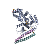
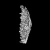
| Title | Structure of the TSC:WIPI3 lysosomal recruitment complex | ||||||||||||
 Components Components |
| ||||||||||||
 Keywords Keywords | ONCOPROTEIN / Tuberous sclerosis complex / tumour suppressor / GTPase-activating proteins (GAP) / TSC-mTORC pathway | ||||||||||||
| Function / homology |  Function and homology information Function and homology informationTSC1-TSC2 complex binding / TSC1-TSC2 complex / Inhibition of TSC complex formation by AKT (PKB) / memory T cell differentiation / regulation of insulin receptor signaling pathway / cellular response to decreased oxygen levels / glycophagy / nucleophagy / negative regulation of cilium assembly / protein localization to phagophore assembly site ...TSC1-TSC2 complex binding / TSC1-TSC2 complex / Inhibition of TSC complex formation by AKT (PKB) / memory T cell differentiation / regulation of insulin receptor signaling pathway / cellular response to decreased oxygen levels / glycophagy / nucleophagy / negative regulation of cilium assembly / protein localization to phagophore assembly site / regulation of cell-matrix adhesion / negative regulation of ATP-dependent activity / phagophore assembly site membrane / activation of GTPase activity / cardiac muscle cell differentiation / response to growth factor / ATPase inhibitor activity / Energy dependent regulation of mTOR by LKB1-AMPK / autophagy of mitochondrion / phosphatidylinositol-3-phosphate binding / pexophagy / regulation of stress fiber assembly / negative regulation of cell size / negative regulation of TOR signaling / regulation of small GTPase mediated signal transduction / phagophore assembly site / TBC/RABGAPs / phosphatidylinositol-3,5-bisphosphate binding / AKT phosphorylates targets in the cytosol / cell projection organization / anoikis / : / negative regulation of macroautophagy / positive chemotaxis / Macroautophagy / Constitutive Signaling by AKT1 E17K in Cancer / negative regulation of mitophagy / ciliary transition zone / positive regulation of macroautophagy / regulation of endocytosis / negative regulation of Wnt signaling pathway / associative learning / positive regulation of focal adhesion assembly / positive regulation of GTPase activity / protein folding chaperone complex / phosphatase binding / autophagosome assembly / adult locomotory behavior / vesicle-mediated transport / negative regulation of TORC1 signaling / myelination / negative regulation of insulin receptor signaling pathway / protein folding chaperone / lipid droplet / Hsp70 protein binding / positive regulation of protein ubiquitination / cell-matrix adhesion / negative regulation of phosphatidylinositol 3-kinase/protein kinase B signal transduction / cellular response to starvation / kidney development / neural tube closure / potassium ion transport / GTPase activator activity / hippocampus development / TP53 Regulates Metabolic Genes / cerebral cortex development / Hsp90 protein binding / response to insulin / synapse organization / protein import into nucleus / small GTPase binding / endocytosis / intracellular protein localization / heart development / lamellipodium / protein-folding chaperone binding / cytoplasmic vesicle / cell cortex / adaptive immune response / protein-macromolecule adaptor activity / cell population proliferation / lysosome / regulation of cell cycle / ciliary basal body / postsynaptic density / protein stabilization / negative regulation of cell population proliferation / lysosomal membrane / perinuclear region of cytoplasm / Golgi apparatus / protein homodimerization activity / protein-containing complex / membrane / nucleus / plasma membrane / cytosol / cytoplasm Similarity search - Function | ||||||||||||
| Biological species |  Homo sapiens (human) Homo sapiens (human) | ||||||||||||
| Method | ELECTRON MICROSCOPY / single particle reconstruction / cryo EM / Resolution: 2.9 Å | ||||||||||||
 Authors Authors | Bayly-Jones, C. / Lupton, C.J. / D'Andrea, L. / Ellisdon, A.M. | ||||||||||||
| Funding support |  United States, United States,  Australia, 3items Australia, 3items
| ||||||||||||
 Citation Citation | Journal: Sci Adv / Year: 2024 Title: Structure of the human TSC:WIPI3 lysosomal recruitment complex. Authors: Charles Bayly-Jones / Christopher J Lupton / Laura D'Andrea / Yong-Gang Chang / Gareth D Jones / Joel R Steele / Hari Venugopal / Ralf B Schittenhelm / Michelle L Halls / Andrew M Ellisdon / Abstract: Tuberous sclerosis complex (TSC) is targeted to the lysosomal membrane, where it hydrolyzes RAS homolog-mTORC1 binding (RHEB) from its GTP-bound to GDP-bound state, inhibiting mechanistic target of ...Tuberous sclerosis complex (TSC) is targeted to the lysosomal membrane, where it hydrolyzes RAS homolog-mTORC1 binding (RHEB) from its GTP-bound to GDP-bound state, inhibiting mechanistic target of rapamycin complex 1 (mTORC1). Loss-of-function mutations in TSC cause TSC disease, marked by excessive tumor growth. Here, we overcome a high degree of continuous conformational heterogeneity to determine the 2.8-Å cryo-electron microscopy (cryo-EM) structure of the complete human TSC in complex with the lysosomal recruitment factor WD repeat domain phosphoinositide-interacting protein 3 (WIPI3). We discover a previously undetected amino-terminal TSC1 HEAT repeat dimer that clamps onto a single TSC wing and forms a phosphatidylinositol phosphate (PIP)-binding pocket, which specifically binds monophosphorylated PIPs. These structural advances provide a model by which WIPI3 and PIP-signaling networks coordinate to recruit TSC to the lysosomal membrane to inhibit mTORC1. The high-resolution TSC structure reveals previously unrecognized mutational hotspots and uncovers crucial insights into the mechanisms of TSC dysregulation in disease. | ||||||||||||
| History |
|
- Structure visualization
| Structure viewer | Molecule: MolmilJmol/JSmol |
|---|
- Downloads & links
-Download
| PDBx/mmCIF format | 9ce3.cif.gz | 900.2 KB | Display | PDBx/mmCIF format |
|---|---|---|---|---|
| PDB format | pdb9ce3.ent.gz | 709.3 KB | Display | PDB format |
| PDBx/mmJSON format | 9ce3.json.gz | Tree view | PDBx/mmJSON format | |
| Others |  Other downloads Other downloads |
-Validation report
| Arichive directory | https://data.pdbj.org/pub/pdb/validation_reports/ce/9ce3ftp://data.pdbj.org/pub/pdb/validation_reports/ce/9ce3 | HTTPS FTP |
|---|
-Related structure data
| Related structure data |  45492MC  9c9iC M: map data used to model this data C: citing same article ( |
|---|---|
| Similar structure data |


-Links
 PDBj
PDBj


- Assembly
| Deposited unit | 
|
|---|---|
| 1 |
|
-Components
| #1: Protein | Mass: 199339.000 Da / Num. of mol.: 2 Source method: isolated from a genetically manipulated source Source: (gene. exp.) Homo sapiens (human) / Gene: TSC2, TSC4 / Production host: Homo sapiens (human) / References: UniProt: P49815#2: Protein | Mass: 133001.609 Da / Num. of mol.: 2 Source method: isolated from a genetically manipulated source Source: (gene. exp.) Homo sapiens (human) / Gene: TSC1, KIAA0243, TSC / Plasmid: pRK7 / Cell line (production host): Expi HEK293 / Production host: Homo sapiens (human) / References: UniProt: Q92574#3: Protein | | Mass: 35016.508 Da / Num. of mol.: 1 Source method: isolated from a genetically manipulated source Source: (gene. exp.) Homo sapiens (human) / Gene: TBC1D7, TBC7, HSPC239 / Production host: Homo sapiens (human) / References: UniProt: Q9P0N9#4: Protein | | Mass: 35222.359 Da / Num. of mol.: 1 Source method: isolated from a genetically manipulated source Source: (gene. exp.) Homo sapiens (human) / Gene: WDR45B, WDR45L, WIPI3 / Production host: Homo sapiens (human) / References: UniProt: Q5MNZ6#5: Protein/peptide | Mass: 1039.273 Da / Num. of mol.: 2 / Source method: isolated from a natural source / Source: (natural) Homo sapiens (human)Has protein modification | N | |
|---|
-Experimental details
-Experiment
| Experiment | Method: ELECTRON MICROSCOPY |
|---|---|
| EM experiment | Aggregation state: PARTICLE / 3D reconstruction method: single particle reconstruction |
- Sample preparation
| Component | Name: TSC:WIPI3 lysosomal recruitment complex (composite map) Type: COMPLEX / Entity ID: all / Source: RECOMBINANT | ||||||||||||||||||||
|---|---|---|---|---|---|---|---|---|---|---|---|---|---|---|---|---|---|---|---|---|---|
| Molecular weight | Value: 0.733 MDa / Experimental value: YES | ||||||||||||||||||||
| Source (natural) | Organism: Homo sapiens (human) | ||||||||||||||||||||
| Source (recombinant) | Organism: Homo sapiens (human) / Cell: Expi HEK293 | ||||||||||||||||||||
| Buffer solution | pH: 7.6 / Details: 20 mM HEPES (pH 7.6), 250 mM NaCl, 2 mM DTT | ||||||||||||||||||||
| Buffer component |
| ||||||||||||||||||||
| Specimen | Conc.: 1.4 mg/ml / Embedding applied: NO / Shadowing applied: NO / Staining applied: NO / Vitrification applied: YES | ||||||||||||||||||||
| Specimen support | Grid material: COPPER / Grid mesh size: 200 divisions/in. / Grid type: Quantifoil R2/2 | ||||||||||||||||||||
| Vitrification | Instrument: FEI VITROBOT MARK IV / Cryogen name: ETHANE / Humidity: 100 % / Chamber temperature: 277.15 K |
- Electron microscopy imaging
| Experimental equipment |  Model: Titan Krios / Image courtesy: FEI Company |
|---|---|
| Microscopy | Model: FEI TITAN KRIOS |
| Electron gun | Electron source:  FIELD EMISSION GUN / Accelerating voltage: 300 kV / Illumination mode: FLOOD BEAM FIELD EMISSION GUN / Accelerating voltage: 300 kV / Illumination mode: FLOOD BEAM |
| Electron lens | Mode: BRIGHT FIELD / Nominal magnification: 105000 X / Nominal defocus max: 3000 nm / Nominal defocus min: 500 nm / Cs: 2.7 mm / C2 aperture diameter: 50.01 µm / Alignment procedure: COMA FREE |
| Specimen holder | Cryogen: NITROGEN / Specimen holder model: FEI TITAN KRIOS AUTOGRID HOLDER |
| Image recording | Average exposure time: 3.71 sec. / Electron dose: 46.54 e/Å2 / Film or detector model: GATAN K3 BIOQUANTUM (6k x 4k) / Num. of grids imaged: 1 / Num. of real images: 12807 |
- Processing
| EM software |
| ||||||||||||||||||||||||||||||||||||||||||||||||||||||||
|---|---|---|---|---|---|---|---|---|---|---|---|---|---|---|---|---|---|---|---|---|---|---|---|---|---|---|---|---|---|---|---|---|---|---|---|---|---|---|---|---|---|---|---|---|---|---|---|---|---|---|---|---|---|---|---|---|---|
| CTF correction | Type: PHASE FLIPPING AND AMPLITUDE CORRECTION | ||||||||||||||||||||||||||||||||||||||||||||||||||||||||
| Particle selection | Num. of particles selected: 1176292 / Details: Template picking, blob picking, TOPAZ, crYOLO | ||||||||||||||||||||||||||||||||||||||||||||||||||||||||
| Symmetry | Point symmetry: C1 (asymmetric) | ||||||||||||||||||||||||||||||||||||||||||||||||||||||||
| 3D reconstruction | Resolution: 2.9 Å / Resolution method: FSC 0.143 CUT-OFF / Num. of particles: 200000 / Algorithm: FOURIER SPACE Details: The global resolution of this composite is estimated based on the voxel average of focused reconstructions. Symmetry type: POINT | ||||||||||||||||||||||||||||||||||||||||||||||||||||||||
| Atomic model building | B value: 96.4 / Protocol: OTHER / Space: REAL / Target criteria: Cross-correlation coefficient | ||||||||||||||||||||||||||||||||||||||||||||||||||||||||
| Atomic model building | 3D fitting-ID: 1
| ||||||||||||||||||||||||||||||||||||||||||||||||||||||||
| Refine LS restraints |
|