Movie
Movie Controller

[English] 日本語
 Yorodumi
- EMDB-45529: The WIPI3:TSC lysosomal docking complex (focused reconstruction; ... -
+ Open data

- Basic information
| Entry |  | ||||||||||||
|---|---|---|---|---|---|---|---|---|---|---|---|---|---|
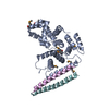
| Title | The WIPI3:TSC lysosomal docking complex (focused reconstruction; WIPI3 TSC2) | ||||||||||||
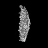
 Map data Map data | Raw final map | ||||||||||||
 Sample Sample |
| ||||||||||||
 Keywords Keywords | Tuberous sclerosis complex / tumour suppressor / GTPase-activating proteins (GAP) / TSC-mTORC pathway / ONCOPROTEIN | ||||||||||||
| Biological species |  Homo sapiens (human) Homo sapiens (human) | ||||||||||||
| Method | single particle reconstruction / cryo EM / Resolution: 3.86 Å | ||||||||||||
 Authors Authors | Bayly-Jones C / Lupton CJ / D'Andrea L / Ellisdon AM | ||||||||||||
| Funding support |  United States, United States,  Australia, 3 items Australia, 3 items
| ||||||||||||
 Citation Citation | Journal: Sci Adv / Year: 2024 Title: Structure of the human TSC:WIPI3 lysosomal recruitment complex. Authors: Charles Bayly-Jones / Christopher J Lupton / Laura D'Andrea / Yong-Gang Chang / Gareth D Jones / Joel R Steele / Hari Venugopal / Ralf B Schittenhelm / Michelle L Halls / Andrew M Ellisdon / Abstract: Tuberous sclerosis complex (TSC) is targeted to the lysosomal membrane, where it hydrolyzes RAS homolog-mTORC1 binding (RHEB) from its GTP-bound to GDP-bound state, inhibiting mechanistic target of ...Tuberous sclerosis complex (TSC) is targeted to the lysosomal membrane, where it hydrolyzes RAS homolog-mTORC1 binding (RHEB) from its GTP-bound to GDP-bound state, inhibiting mechanistic target of rapamycin complex 1 (mTORC1). Loss-of-function mutations in TSC cause TSC disease, marked by excessive tumor growth. Here, we overcome a high degree of continuous conformational heterogeneity to determine the 2.8-Å cryo-electron microscopy (cryo-EM) structure of the complete human TSC in complex with the lysosomal recruitment factor WD repeat domain phosphoinositide-interacting protein 3 (WIPI3). We discover a previously undetected amino-terminal TSC1 HEAT repeat dimer that clamps onto a single TSC wing and forms a phosphatidylinositol phosphate (PIP)-binding pocket, which specifically binds monophosphorylated PIPs. These structural advances provide a model by which WIPI3 and PIP-signaling networks coordinate to recruit TSC to the lysosomal membrane to inhibit mTORC1. The high-resolution TSC structure reveals previously unrecognized mutational hotspots and uncovers crucial insights into the mechanisms of TSC dysregulation in disease. | ||||||||||||
| History |
|
- Structure visualization
| Supplemental images |
|---|

- Downloads & links
-EMDB archive
| Map data | emd_45529.map.gz | 32.3 MB |  EMDB map data format EMDB map data format | |
|---|---|---|---|---|
| Header (meta data) | emd-45529-v30.xmlemd-45529.xml | 23.6 KB 23.6 KB | Display Display | EMDB header |
| FSC (resolution estimation) | emd_45529_fsc.xml | 8.4 KB | Display | FSC data file |
| Images |  emd_45529.png emd_45529.png | 59.7 KB | ||
| Masks | emd_45529_msk_1.map | 64 MB | Mask map | |
| Filedesc metadata | emd-45529.cif.gz | 5.2 KB | ||
| Others | emd_45529_additional_1.map.gzemd_45529_half_map_1.map.gzemd_45529_half_map_2.map.gz | 53.3 MB 59.4 MB 59.4 MB | ||
| Archive directory |  http://ftp.pdbj.org/pub/emdb/structures/EMD-45529ftp://ftp.pdbj.org/pub/emdb/structures/EMD-45529 http://ftp.pdbj.org/pub/emdb/structures/EMD-45529ftp://ftp.pdbj.org/pub/emdb/structures/EMD-45529 | HTTPS FTP |
-Related structure data


-Links
| EMDB pages | EMDB (EBI/PDBe) / EMDataResource |
|---|
-Map
| File | Download / File: emd_45529.map.gz / Format: CCP4 / Size: 64 MB / Type: IMAGE STORED AS FLOATING POINT NUMBER (4 BYTES) | ||||||||||||||||||||||||||||||||||||
|---|---|---|---|---|---|---|---|---|---|---|---|---|---|---|---|---|---|---|---|---|---|---|---|---|---|---|---|---|---|---|---|---|---|---|---|---|---|
| Annotation | Raw final map | ||||||||||||||||||||||||||||||||||||
| Projections & slices | Image control
Images are generated by Spider. | ||||||||||||||||||||||||||||||||||||
| Voxel size | X=Y=Z: 1.1714 Å | ||||||||||||||||||||||||||||||||||||
| Density |
| ||||||||||||||||||||||||||||||||||||
| Symmetry | Space group: 1 | ||||||||||||||||||||||||||||||||||||
| Details | EMDB XML:
|
 Z (Sec.)
 Y (Row.)
 X (Col.)


-Supplemental data
-Mask #1
| File | emd_45529_msk_1.map | ||||||||||||
|---|---|---|---|---|---|---|---|---|---|---|---|---|---|
| Projections & Slices |
| ||||||||||||
| Density Histograms |


-Additional map: Sharpened map
| File | emd_45529_additional_1.map | ||||||||||||
|---|---|---|---|---|---|---|---|---|---|---|---|---|---|
| Annotation | Sharpened map | ||||||||||||
| Projections & Slices |
| ||||||||||||
| Density Histograms |


-Half map: Unfiltered half map (A)
| File | emd_45529_half_map_1.map | ||||||||||||
|---|---|---|---|---|---|---|---|---|---|---|---|---|---|
| Annotation | Unfiltered half map (A) | ||||||||||||
| Projections & Slices |
| ||||||||||||
| Density Histograms |


-Half map: Unfiltered half map (B)
| File | emd_45529_half_map_2.map | ||||||||||||
|---|---|---|---|---|---|---|---|---|---|---|---|---|---|
| Annotation | Unfiltered half map (B) | ||||||||||||
| Projections & Slices |
| ||||||||||||
| Density Histograms |


- Sample components
-Entire : TSC:WIPI3 lysosomal recruitment complex
| Entire | Name: TSC:WIPI3 lysosomal recruitment complex |
|---|---|
| Components |
|
-Supramolecule #1: TSC:WIPI3 lysosomal recruitment complex
| Supramolecule | Name: TSC:WIPI3 lysosomal recruitment complex / type: complex / ID: 1 / Parent: 0 / Macromolecule list: #1-#7 |
|---|---|
| Source (natural) | Organism: Homo sapiens (human) |
| Molecular weight | Theoretical: 733 KDa |
-Experimental details
-Structure determination
| Method | cryo EM |
|---|---|
 Processing Processing | single particle reconstruction |
| Aggregation state | particle |
-Sample preparation
| Concentration | 1.4 mg/mL | ||||||||||||
|---|---|---|---|---|---|---|---|---|---|---|---|---|---|
| Buffer | pH: 7.6 Component:
Details: 20 mM HEPES (pH 7.6), 250 mM NaCl, 2 mM DTT | ||||||||||||
| Grid | Model: Quantifoil R1.2/1.3 / Material: COPPER / Mesh: 200 / Support film - Material: CARBON / Support film - topology: HOLEY | ||||||||||||
| Vitrification | Cryogen name: ETHANE / Chamber humidity: 100 % / Chamber temperature: 277.15 K / Instrument: FEI VITROBOT MARK IV |
- Electron microscopy
| Microscope | FEI TITAN KRIOS |
|---|---|
| Image recording | Film or detector model: GATAN K3 BIOQUANTUM (6k x 4k) / Number grids imaged: 1 / Number real images: 12807 / Average exposure time: 3.71 sec. / Average electron dose: 46.54 e/Å2 |
| Electron beam | Acceleration voltage: 300 kV / Electron source:  FIELD EMISSION GUN FIELD EMISSION GUN |
| Electron optics | C2 aperture diameter: 50.01 µm / Illumination mode: FLOOD BEAM / Imaging mode: BRIGHT FIELD / Cs: 2.7 mm / Nominal defocus max: 3.0 µm / Nominal defocus min: 0.5 µm / Nominal magnification: 105000 |
| Sample stage | Specimen holder model: FEI TITAN KRIOS AUTOGRID HOLDER / Cooling holder cryogen: NITROGEN |
| Experimental equipment |  Model: Titan Krios / Image courtesy: FEI Company |
+Image processing

-Atomic model buiding 1
| Initial model |
| ||||||||||
|---|---|---|---|---|---|---|---|---|---|---|---|
| Refinement | Space: REAL / Protocol: OTHER / Overall B value: 96.4 / Target criteria: Cross-correlation coefficient |