[English] 日本語
 Yorodumi
- PDB-9c67: cryoEM structure of CRISPR associated effector, CARF-Adenosine de... -
+ Open data
- Basic information
| Entry | Database: PDB / ID: 9c67 | ||||||
|---|---|---|---|---|---|---|---|
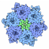
| Title | cryoEM structure of CRISPR associated effector, CARF-Adenosine deaminase 1, Cad1, in apo form | ||||||
 Components Components | Adenosine deaminase domain-containing protein | ||||||
 Keywords Keywords | ANTIVIRAL PROTEIN / Antiphage defense / CRISPR / Deamination / CARF-Adenosine deaminase | ||||||
| Function / homology |  Function and homology information Function and homology informationinosine biosynthetic process / adenosine deaminase / hypoxanthine salvage / adenosine catabolic process / adenosine deaminase activity / cytosol Similarity search - Function | ||||||
| Biological species |  Bacteroidales bacterium (bacteria) Bacteroidales bacterium (bacteria) | ||||||
| Method | ELECTRON MICROSCOPY / single particle reconstruction / cryo EM / Resolution: 3.6 Å | ||||||
 Authors Authors | Majumder, P. / Patel, D.J. | ||||||
| Funding support |  United States, 1items United States, 1items
| ||||||
 Citation Citation |  Journal: Cell / Year: 2024 Journal: Cell / Year: 2024Title: The CRISPR-associated adenosine deaminase Cad1 converts ATP to ITP to provide antiviral immunity. Authors: Christian F Baca / Puja Majumder / James H Hickling / Linzhi Ye / Marianna Teplova / Sean F Brady / Dinshaw J Patel / Luciano A Marraffini /  Abstract: Type III CRISPR systems provide immunity against genetic invaders through the production of cyclic oligo-adenylate (cA) molecules that activate effector proteins that contain CRISPR-associated ...Type III CRISPR systems provide immunity against genetic invaders through the production of cyclic oligo-adenylate (cA) molecules that activate effector proteins that contain CRISPR-associated Rossman fold (CARF) domains. Here, we characterized the function and structure of an effector in which the CARF domain is fused to an adenosine deaminase domain, CRISPR-associated adenosine deaminase 1 (Cad1). We show that upon binding of cA or cA to its CARF domain, Cad1 converts ATP to ITP, both in vivo and in vitro. Cryoelectron microscopy (cryo-EM) structural studies on full-length Cad1 reveal an hexameric assembly composed of a trimer of dimers, with bound ATP at inter-domain sites required for activity and ATP/ITP within deaminase active sites. Upon synthesis of cA during phage infection, Cad1 activation leads to a growth arrest of the host that prevents viral propagation. Our findings reveal that CRISPR-Cas systems employ a wide range of molecular mechanisms beyond nucleic acid degradation to provide adaptive immunity in prokaryotes. | ||||||
| History |
|
- Structure visualization
| Structure viewer | Molecule:  Molmil Molmil Jmol/JSmol Jmol/JSmol |
|---|
- Downloads & links
- Download
| PDBx/mmCIF format |  9c67.cif.gz 9c67.cif.gz | 573.1 KB | Display |  PDBx/mmCIF format PDBx/mmCIF format |
|---|---|---|---|---|
| PDB format |  pdb9c67.ent.gz pdb9c67.ent.gz | 480.8 KB | Display |  PDB format PDB format |
| PDBx/mmJSON format |  9c67.json.gz 9c67.json.gz | Tree view |  PDBx/mmJSON format PDBx/mmJSON format | |
| Others |  Other downloads Other downloads |
-Validation report
| Arichive directory |  https://data.pdbj.org/pub/pdb/validation_reports/c6/9c67 https://data.pdbj.org/pub/pdb/validation_reports/c6/9c67 ftp://data.pdbj.org/pub/pdb/validation_reports/c6/9c67 ftp://data.pdbj.org/pub/pdb/validation_reports/c6/9c67 | HTTPS FTP |
|---|
-Related structure data
| Related structure data |  45241MC  9c68C  9c69C  9c6aC  9c6cC  9c6fC  9c77C  9cdbC M: map data used to model this data C: citing same article ( |
|---|---|
| Similar structure data | Similarity search - Function & homology  F&H Search F&H Search |
- Links
Links
- Assembly
| Deposited unit | 
|
|---|---|
| 1 |
|
- Components
| #1: Protein | Mass: 67264.945 Da / Num. of mol.: 6 Source method: isolated from a genetically manipulated source Source: (gene. exp.)  Bacteroidales bacterium (bacteria) / Gene: DCM62_02910 / Production host: Bacteroidales bacterium (bacteria) / Gene: DCM62_02910 / Production host:  #2: Chemical | ChemComp-MG / Has ligand of interest | Y | Has protein modification | N | |
|---|
-Experimental details
-Experiment
| Experiment | Method: ELECTRON MICROSCOPY |
|---|---|
| EM experiment | Aggregation state: PARTICLE / 3D reconstruction method: single particle reconstruction |
- Sample preparation
| Component | Name: apo Cad1 (CRISPR associated Adenosine deaminase 1) full length protein Type: COMPLEX / Details: It forms a trimer of dimers / Entity ID: #1 / Source: RECOMBINANT |
|---|---|
| Molecular weight | Value: 0.4 MDa / Experimental value: YES |
| Source (natural) | Organism:  Bacteriodale bacterium (bacteria) Bacteriodale bacterium (bacteria) |
| Source (recombinant) | Organism:  |
| Buffer solution | pH: 8 Details: 25 mM Hepes pH 8, 200 mM NaCl, 2 mM Beta-mercaptoethanol and 5 % glycerol |
| Specimen | Conc.: 1.7 mg/ml / Embedding applied: NO / Shadowing applied: NO / Staining applied: NO / Vitrification applied: YES |
| Specimen support | Details: Glow discharge 1 min, 15 mA 100 % humidity, wait time 12 s, 2.5 s blot time and 0 blot force at 4 degree C temperature Grid material: GOLD / Grid mesh size: 300 divisions/in. / Grid type: UltrAuFoil R1.2/1.3 |
| Vitrification | Instrument: FEI VITROBOT MARK IV / Cryogen name: ETHANE / Humidity: 100 % / Chamber temperature: 277 K |
- Electron microscopy imaging
| Experimental equipment |  Model: Titan Krios / Image courtesy: FEI Company |
|---|---|
| Microscopy | Model: FEI TITAN KRIOS |
| Electron gun | Electron source:  FIELD EMISSION GUN / Accelerating voltage: 300 kV / Illumination mode: FLOOD BEAM FIELD EMISSION GUN / Accelerating voltage: 300 kV / Illumination mode: FLOOD BEAM |
| Electron lens | Mode: BRIGHT FIELD / Nominal defocus max: 2500 nm / Nominal defocus min: 800 nm / Cs: 2.7 mm |
| Specimen holder | Cryogen: NITROGEN |
| Image recording | Electron dose: 53 e/Å2 / Detector mode: SUPER-RESOLUTION / Film or detector model: GATAN K3 (6k x 4k) |
| EM imaging optics | Energyfilter slit width: 20 eV |
- Processing
| EM software |
| ||||||||||||||||||||||||
|---|---|---|---|---|---|---|---|---|---|---|---|---|---|---|---|---|---|---|---|---|---|---|---|---|---|
| CTF correction | Type: NONE | ||||||||||||||||||||||||
| 3D reconstruction | Resolution: 3.6 Å / Resolution method: FSC 0.143 CUT-OFF / Num. of particles: 110836 Details: Half maps were generated by Non-uniform refinement programme using cryoSparc Num. of class averages: 1 / Symmetry type: POINT | ||||||||||||||||||||||||
| Atomic model building | Protocol: RIGID BODY FIT / Space: REAL | ||||||||||||||||||||||||
| Atomic model building | Source name: AlphaFold / Type: in silico model | ||||||||||||||||||||||||
| Refine LS restraints |
|
 Movie
Movie Controller


 PDBj
PDBj


