[English] 日本語
 Yorodumi
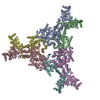
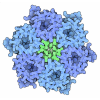
- EMDB-45466: CryoEM structure of CRISPR associated effector, CARF-Adenosine de... -
+ Open data
- Basic information
| Entry |  | |||||||||
|---|---|---|---|---|---|---|---|---|---|---|
| Title | CryoEM structure of CRISPR associated effector, CARF-Adenosine deaminase 1, Cad1, in cA6 (partial density) bound form with ATP (partial density). | |||||||||
 Map data Map data | structure of CRISPR associated effector, CARF-Adenosine deaminase 1, Cad1, in cA6 (partial density) bound form with ATP (partial density). | |||||||||
 Sample Sample |
| |||||||||
 Keywords Keywords | Antiphage defense / CRISPR / Deamination / CARF-Adenosine deaminase / ATP / ANTIVIRAL PROTEIN / ANTIVIRAL PROTEIN-RNA complex | |||||||||
| Function / homology |  Function and homology information Function and homology informationinosine biosynthetic process / adenosine deaminase / hypoxanthine salvage / adenosine deaminase activity / adenosine catabolic process / cytosol Similarity search - Function | |||||||||
| Biological species |  bacteroidale bacteria (bacteria) / bacteroidale bacteria (bacteria) /  Bacteroidales bacterium (bacteria) Bacteroidales bacterium (bacteria) | |||||||||
| Method | single particle reconstruction / cryo EM / Resolution: 3.6 Å | |||||||||
 Authors Authors | Majumder P / Patel DJ | |||||||||
| Funding support |  United States, 1 items United States, 1 items
| |||||||||
 Citation Citation |  Journal: Cell / Year: 2024 Journal: Cell / Year: 2024Title: The CRISPR-associated adenosine deaminase Cad1 converts ATP to ITP to provide antiviral immunity. Authors: Christian F Baca / Puja Majumder / James H Hickling / Linzhi Ye / Marianna Teplova / Sean F Brady / Dinshaw J Patel / Luciano A Marraffini /  Abstract: Type III CRISPR systems provide immunity against genetic invaders through the production of cyclic oligo-adenylate (cA) molecules that activate effector proteins that contain CRISPR-associated ...Type III CRISPR systems provide immunity against genetic invaders through the production of cyclic oligo-adenylate (cA) molecules that activate effector proteins that contain CRISPR-associated Rossman fold (CARF) domains. Here, we characterized the function and structure of an effector in which the CARF domain is fused to an adenosine deaminase domain, CRISPR-associated adenosine deaminase 1 (Cad1). We show that upon binding of cA or cA to its CARF domain, Cad1 converts ATP to ITP, both in vivo and in vitro. Cryoelectron microscopy (cryo-EM) structural studies on full-length Cad1 reveal an hexameric assembly composed of a trimer of dimers, with bound ATP at inter-domain sites required for activity and ATP/ITP within deaminase active sites. Upon synthesis of cA during phage infection, Cad1 activation leads to a growth arrest of the host that prevents viral propagation. Our findings reveal that CRISPR-Cas systems employ a wide range of molecular mechanisms beyond nucleic acid degradation to provide adaptive immunity in prokaryotes. | |||||||||
| History |
|
- Structure visualization
| Supplemental images |
|---|
- Downloads & links
-EMDB archive
| Map data |  emd_45466.map.gz emd_45466.map.gz | 254.9 MB |  EMDB map data format EMDB map data format | |
|---|---|---|---|---|
| Header (meta data) |  emd-45466-v30.xml emd-45466-v30.xml emd-45466.xml emd-45466.xml | 17.4 KB 17.4 KB | Display Display |  EMDB header EMDB header |
| FSC (resolution estimation) |  emd_45466_fsc.xml emd_45466_fsc.xml | 16.9 KB | Display |  FSC data file FSC data file |
| Images |  emd_45466.png emd_45466.png | 68.2 KB | ||
| Filedesc metadata |  emd-45466.cif.gz emd-45466.cif.gz | 6.4 KB | ||
| Others |  emd_45466_half_map_1.map.gz emd_45466_half_map_1.map.gz emd_45466_half_map_2.map.gz emd_45466_half_map_2.map.gz | 475.4 MB 475.4 MB | ||
| Archive directory |  http://ftp.pdbj.org/pub/emdb/structures/EMD-45466 http://ftp.pdbj.org/pub/emdb/structures/EMD-45466 ftp://ftp.pdbj.org/pub/emdb/structures/EMD-45466 ftp://ftp.pdbj.org/pub/emdb/structures/EMD-45466 | HTTPS FTP |
-Validation report
| Summary document |  emd_45466_validation.pdf.gz emd_45466_validation.pdf.gz | 830.8 KB | Display |  EMDB validaton report EMDB validaton report |
|---|---|---|---|---|
| Full document |  emd_45466_full_validation.pdf.gz emd_45466_full_validation.pdf.gz | 830.4 KB | Display | |
| Data in XML |  emd_45466_validation.xml.gz emd_45466_validation.xml.gz | 26.4 KB | Display | |
| Data in CIF |  emd_45466_validation.cif.gz emd_45466_validation.cif.gz | 34.8 KB | Display | |
| Arichive directory |  https://ftp.pdbj.org/pub/emdb/validation_reports/EMD-45466 https://ftp.pdbj.org/pub/emdb/validation_reports/EMD-45466 ftp://ftp.pdbj.org/pub/emdb/validation_reports/EMD-45466 ftp://ftp.pdbj.org/pub/emdb/validation_reports/EMD-45466 | HTTPS FTP |
-Related structure data
| Related structure data |  9cdbMC  9c67C  9c68C  9c69C  9c6aC  9c6cC  9c6fC  9c77C M: atomic model generated by this map C: citing same article ( |
|---|---|
| Similar structure data | Similarity search - Function & homology  F&H Search F&H Search |
- Links
Links
| EMDB pages |  EMDB (EBI/PDBe) / EMDB (EBI/PDBe) /  EMDataResource EMDataResource |
|---|---|
| Related items in Molecule of the Month |
- Map
Map
| File |  Download / File: emd_45466.map.gz / Format: CCP4 / Size: 512 MB / Type: IMAGE STORED AS FLOATING POINT NUMBER (4 BYTES) Download / File: emd_45466.map.gz / Format: CCP4 / Size: 512 MB / Type: IMAGE STORED AS FLOATING POINT NUMBER (4 BYTES) | ||||||||||||||||||||||||||||||||||||
|---|---|---|---|---|---|---|---|---|---|---|---|---|---|---|---|---|---|---|---|---|---|---|---|---|---|---|---|---|---|---|---|---|---|---|---|---|---|
| Annotation | structure of CRISPR associated effector, CARF-Adenosine deaminase 1, Cad1, in cA6 (partial density) bound form with ATP (partial density). | ||||||||||||||||||||||||||||||||||||
| Projections & slices | Image control
Images are generated by Spider. | ||||||||||||||||||||||||||||||||||||
| Voxel size | X=Y=Z: 0.809 Å | ||||||||||||||||||||||||||||||||||||
| Density |
| ||||||||||||||||||||||||||||||||||||
| Symmetry | Space group: 1 | ||||||||||||||||||||||||||||||||||||
| Details | EMDB XML:
|
-Supplemental data
-Half map: Half Map A
| File | emd_45466_half_map_1.map | ||||||||||||
|---|---|---|---|---|---|---|---|---|---|---|---|---|---|
| Annotation | Half Map A | ||||||||||||
| Projections & Slices |
| ||||||||||||
| Density Histograms |
-Half map: Half Map B
| File | emd_45466_half_map_2.map | ||||||||||||
|---|---|---|---|---|---|---|---|---|---|---|---|---|---|
| Annotation | Half Map B | ||||||||||||
| Projections & Slices |
| ||||||||||||
| Density Histograms |
- Sample components
-Entire : cA6 (partial density)-Cad1-ATP (partial density)
| Entire | Name: cA6 (partial density)-Cad1-ATP (partial density) |
|---|---|
| Components |
|
-Supramolecule #1: cA6 (partial density)-Cad1-ATP (partial density)
| Supramolecule | Name: cA6 (partial density)-Cad1-ATP (partial density) / type: complex / ID: 1 / Parent: 0 / Macromolecule list: #1-#2 Details: Cad1 forms a trimer of dimers. In this structure partial density of bound cA6 is visible. ATP is present. |
|---|---|
| Source (natural) | Organism:  bacteroidale bacteria (bacteria) bacteroidale bacteria (bacteria) |
| Molecular weight | Theoretical: 400 KDa |
-Macromolecule #1: Adenosine deaminase domain-containing protein
| Macromolecule | Name: Adenosine deaminase domain-containing protein / type: protein_or_peptide / ID: 1 / Number of copies: 6 / Enantiomer: LEVO |
|---|---|
| Source (natural) | Organism:  Bacteroidales bacterium (bacteria) Bacteroidales bacterium (bacteria) |
| Molecular weight | Theoretical: 67.264945 KDa |
| Recombinant expression | Organism:  |
| Sequence | String: MSRVLLCSAG HSSMVVPEAF HAVPEGFEEV HVFTTDSEKF NPVVLNDFFH SLPNVRFSIT KCHGLADILN ERDFEFYQEM LWQWYLTKM PDNELPYVCL SGGIKSMSAS LQKAATLFGA QSVFHVLADN NPRNIEEMFD ALQKGQIHFI EMGYEPGWAA L RRLKKILP ...String: MSRVLLCSAG HSSMVVPEAF HAVPEGFEEV HVFTTDSEKF NPVVLNDFFH SLPNVRFSIT KCHGLADILN ERDFEFYQEM LWQWYLTKM PDNELPYVCL SGGIKSMSAS LQKAATLFGA QSVFHVLADN NPRNIEEMFD ALQKGQIHFI EMGYEPGWAA L RRLKKILP INEGCSRDNF KPLISKSIEE ILSNVKIMAS DTGKSNQLPF PSLAILPPIA QQWLQLPLSA NDGAWIQNLP KV DLHCHLG GFATSGSLLD QVRGAASEPD LIDRTFSPQE IAGWPRSHKS ISLDKYMELG NANGSKLLKD KGCLIRQVEL LYQ SLVNDN VAYAEIRCSP NNYADKNKNR SAWVVLQDIN DTFTRLITEA KQKNQFYCHV NLLVIASRKF SGDLSDISKH LALA ITAMQ QGEGVCRIVG VDLAGFENKE TRASYYEHDF KAVHRCGLAV TAHAGENDDP EGIWQAVYSL HARRLGHALN LLEAP DLMR TVIERKIGVE MCPYANYQIK GFAPMPNFSA LYPLKKYLEA GILVSVNTDN IGISGANLSE NLLILADLCP GISRMD VLT IIRNSIETAF ISHDFRMELL KFFDRKIYDV CLISIKN UniProtKB: adenosine deaminase |
-Macromolecule #2: RNA (5'-R(P*AP*AP*AP*AP*AP*A)-3')
| Macromolecule | Name: RNA (5'-R(P*AP*AP*AP*AP*AP*A)-3') / type: rna / ID: 2 / Number of copies: 3 |
|---|---|
| Source (natural) | Organism:  Bacteroidales bacterium (bacteria) Bacteroidales bacterium (bacteria) |
| Molecular weight | Theoretical: 1.930277 KDa |
| Sequence | String: AAAAAA |
-Macromolecule #3: ADENOSINE-5'-TRIPHOSPHATE
| Macromolecule | Name: ADENOSINE-5'-TRIPHOSPHATE / type: ligand / ID: 3 / Number of copies: 12 / Formula: ATP |
|---|---|
| Molecular weight | Theoretical: 507.181 Da |
| Chemical component information |  ChemComp-ATP: |
-Macromolecule #4: MAGNESIUM ION
| Macromolecule | Name: MAGNESIUM ION / type: ligand / ID: 4 / Number of copies: 6 / Formula: MG |
|---|---|
| Molecular weight | Theoretical: 24.305 Da |
-Experimental details
-Structure determination
| Method | cryo EM |
|---|---|
 Processing Processing | single particle reconstruction |
| Aggregation state | particle |
- Sample preparation
| Concentration | 1.7 mg/mL |
|---|---|
| Buffer | pH: 8 Details: 25 mM Hepes pH 8, 500 mM NaCl, 2 mM Beta-Mercaptoethanol and 5 percentage glycerol |
| Grid | Model: UltrAuFoil R1.2/1.3 / Material: GOLD / Mesh: 300 / Pretreatment - Type: GLOW DISCHARGE / Pretreatment - Time: 60 sec. |
| Vitrification | Cryogen name: ETHANE / Chamber humidity: 100 % / Chamber temperature: 277.15 K / Instrument: FEI VITROBOT MARK IV / Details: Blot time 2.5 s, Wait time 12s, Blot force 0.. |
| Details | Cad1 protein mixed with cA6 and ATP. Incubated overnight at 310.15 K. |
- Electron microscopy
| Microscope | FEI TITAN KRIOS |
|---|---|
| Specialist optics | Energy filter - Slit width: 20 eV |
| Image recording | Film or detector model: GATAN K3 (6k x 4k) / Average electron dose: 61.0 e/Å2 |
| Electron beam | Acceleration voltage: 300 kV / Electron source:  FIELD EMISSION GUN FIELD EMISSION GUN |
| Electron optics | Illumination mode: FLOOD BEAM / Imaging mode: BRIGHT FIELD / Cs: 2.7 mm / Nominal defocus max: 2.4 µm / Nominal defocus min: 0.8 µm |
| Sample stage | Cooling holder cryogen: NITROGEN |
| Experimental equipment |  Model: Titan Krios / Image courtesy: FEI Company |
+ Image processing
-Atomic model buiding 1
| Initial model | Chain - Source name: AlphaFold / Chain - Initial model type: in silico model |
|---|---|
| Refinement | Protocol: AB INITIO MODEL |
| Output model |  PDB-9cdb: |
 Movie
Movie Controller


 Z (Sec.)
 Y (Row.)
 X (Col.)


