 Movie
Movie Controller


+ Open data

- Basic information
| Entry | Database: PDB / ID: 9az5 | ||||||
|---|---|---|---|---|---|---|---|
| Title | Mycolicibacterium smegmatis ClpS with bound Mg2+ | ||||||
 Components Components | ATP-dependent Clp protease adapter protein ClpS | ||||||
 Keywords Keywords | PROTEIN BINDING / proteolysis / adaptor | ||||||
| Function / homology | ATP-dependent Clp protease adaptor protein ClpS / Adaptor protein ClpS, core / ATP-dependent Clp protease adaptor protein ClpS / Ribosomal protein L7/L12, C-terminal/adaptor protein ClpS-like / protein catabolic process / peptidase activity / proteolysis / NICKEL (II) ION / ATP-dependent Clp protease adapter protein ClpS Function and homology information Function and homology information | ||||||
| Biological species |  Mycolicibacterium smegmatis MC2 155 (bacteria) Mycolicibacterium smegmatis MC2 155 (bacteria) | ||||||
| Method |  X-RAY DIFFRACTION / MOLECULAR REPLACEMENT / Resolution: 1.47 Å X-RAY DIFFRACTION / MOLECULAR REPLACEMENT / Resolution: 1.47 Å | ||||||
 Authors Authors | Presloid, C.J. / Jiang, J. / Beardslee, P.C. / Anderson, H.R. / Swayne, T.M. / Schmitz, K.R. | ||||||
| Funding support |  United States, 1items United States, 1items
| ||||||
 Citation Citation | Journal: Mol.Microbiol. / Year: 2025 Title: ClpS Directs Degradation of N-Degron Substrates With Primary Destabilizing Residues in Mycolicibacterium smegmatis. Authors: Presloid, C.J. / Jiang, J. / Kandel, P. / Anderson, H.R. / Beardslee, P.C. / Swayne, T.M. / Schmitz, K.R. | ||||||
| History |
|
- Structure visualization
| Structure viewer | Molecule: MolmilJmol/JSmol |
|---|
- Downloads & links
-Download
| PDBx/mmCIF format | 9az5.cif.gz | 114.8 KB | Display | PDBx/mmCIF format |
|---|---|---|---|---|
| PDB format | pdb9az5.ent.gz | 84.9 KB | Display | PDB format |
| PDBx/mmJSON format | 9az5.json.gz | Tree view | PDBx/mmJSON format | |
| Others |  Other downloads Other downloads |
-Validation report
| Arichive directory | https://data.pdbj.org/pub/pdb/validation_reports/az/9az5ftp://data.pdbj.org/pub/pdb/validation_reports/az/9az5 | HTTPS FTP |
|---|
-Related structure data
| Related structure data |  9aynC  9ayoC  9aypC  9b06C 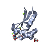 9b10C  9b1pC C: citing same article ( |
|---|---|
| Similar structure data |
-Links
 PDBj
PDBj

- Assembly
| Deposited unit | 
| ||||||||||||
|---|---|---|---|---|---|---|---|---|---|---|---|---|---|
| 1 | 
| ||||||||||||
| 2 | 
| ||||||||||||
| Unit cell |
|
-Components
| #1: Protein | Mass: 9022.167 Da / Num. of mol.: 2 Source method: isolated from a genetically manipulated source Source: (gene. exp.) Mycolicibacterium smegmatis MC2 155 (bacteria)Gene: clpS, MSMEG_4910 / Plasmid: pET22b / Production host: #2: Chemical | ChemComp-MG /   Mass: 24.305 Da / Num. of mol.: 5 / Source method: obtained synthetically / Formula: Mg Mass: 24.305 Da / Num. of mol.: 5 / Source method: obtained synthetically / Formula: Mg#3: Chemical | ChemComp-DMS / |   Mass: 78.133 Da / Num. of mol.: 1 / Source method: obtained synthetically / Formula: C2H6OS / Comment: DMSO, precipitant*YM Mass: 78.133 Da / Num. of mol.: 1 / Source method: obtained synthetically / Formula: C2H6OS / Comment: DMSO, precipitant*YM#4: Chemical | ChemComp-NI / |   Mass: 58.693 Da / Num. of mol.: 1 / Source method: obtained synthetically / Formula: Ni Mass: 58.693 Da / Num. of mol.: 1 / Source method: obtained synthetically / Formula: Ni#5: Water | ChemComp-HOH / |  Mass: 18.015 Da / Num. of mol.: 279 / Source method: isolated from a natural source / Formula: H2O Mass: 18.015 Da / Num. of mol.: 279 / Source method: isolated from a natural source / Formula: H2OHas ligand of interest | N | Has protein modification | N | |
|---|
-Experimental details
-Experiment
| Experiment | Method: X-RAY DIFFRACTION / Number of used crystals: 1 |
|---|
- Sample preparation
| Crystal | Density Matthews: 2.03 Å3/Da / Density % sol: 39.47 % |
|---|---|
| Crystal grow | Temperature: 293 K / Method: vapor diffusion, hanging drop / pH: 7.5 Details: 0.1 M HEPES pH 7.5, 12% PEG3350, 5 mM NiCl2 cryo: 0.1 M HEPES pH 7.5, 40% PEG3350, 5 mM MgCl2 |
-Data collection
| Diffraction | Mean temperature: 100 K / Serial crystal experiment: N |
|---|---|
| Diffraction source | Source: ROTATING ANODE / Type: RIGAKU MICROMAX-007 / Wavelength: 1.54178 Å |
| Detector | Type: RIGAKU SATURN 944 / Detector: CCD / Date: Jan 28, 2017 |
| Radiation | Protocol: SINGLE WAVELENGTH / Monochromatic (M) / Laue (L): M / Scattering type: x-ray |
| Radiation wavelength | Wavelength: 1.54178 Å / Relative weight: 1 |
| Reflection | Resolution: 1.47→30.43 Å / Num. obs: 24656 / % possible obs: 95.9 % / Redundancy: 7.5 % / Biso Wilson estimate: 12.37 Å2 / Rmerge(I) obs: 0.048 / Rpim(I) all: 0.017 / Rrim(I) all: 0.052 / Net I/σ(I): 23.9 |
| Reflection shell | Resolution: 1.47→1.5 Å / Redundancy: 1.7 % / Rmerge(I) obs: 0.509 / Mean I/σ(I) obs: 1.14 / Num. unique obs: 652 / CC1/2: 0.698 / Rpim(I) all: 0.472 / Rrim(I) all: 0.696 |
- Processing
| Software |
| ||||||||||||||||||||||||||||||||||||||||||||||||||||||||||||||||||||||
|---|---|---|---|---|---|---|---|---|---|---|---|---|---|---|---|---|---|---|---|---|---|---|---|---|---|---|---|---|---|---|---|---|---|---|---|---|---|---|---|---|---|---|---|---|---|---|---|---|---|---|---|---|---|---|---|---|---|---|---|---|---|---|---|---|---|---|---|---|---|---|---|
| Refinement | Method to determine structure: MOLECULAR REPLACEMENT / Resolution: 1.47→27.3 Å / SU ML: 0.1742 / Cross valid method: FREE R-VALUE / σ(F): 1.33 / Phase error: 19.6312 Stereochemistry target values: GeoStd + Monomer Library + CDL v1.2
| ||||||||||||||||||||||||||||||||||||||||||||||||||||||||||||||||||||||
| Solvent computation | Shrinkage radii: 0.9 Å / VDW probe radii: 1.11 Å / Solvent model: FLAT BULK SOLVENT MODEL | ||||||||||||||||||||||||||||||||||||||||||||||||||||||||||||||||||||||
| Displacement parameters | Biso mean: 17.83 Å2 | ||||||||||||||||||||||||||||||||||||||||||||||||||||||||||||||||||||||
| Refinement step | Cycle: LAST / Resolution: 1.47→27.3 Å
| ||||||||||||||||||||||||||||||||||||||||||||||||||||||||||||||||||||||
| Refine LS restraints |
| ||||||||||||||||||||||||||||||||||||||||||||||||||||||||||||||||||||||
| LS refinement shell |
|
