Movie
Movie Controller

[English] 日本語
 Yorodumi
- PDB-8zds: Structure of the Salmonella flagellar MS-ring with C11 symmetry a... -
+ Open data

- Basic information
| Entry | Database: PDB / ID: 8zds | |||||||||||||||||||||||||||||||||
|---|---|---|---|---|---|---|---|---|---|---|---|---|---|---|---|---|---|---|---|---|---|---|---|---|---|---|---|---|---|---|---|---|---|---|
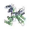
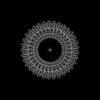
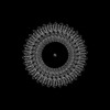
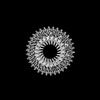
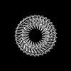
| Title | Structure of the Salmonella flagellar MS-ring with C11 symmetry applied | |||||||||||||||||||||||||||||||||
 Components Components | Flagellar M-ring protein | |||||||||||||||||||||||||||||||||
 Keywords Keywords | MOTOR PROTEIN / Bacterial flagellum / flagellar assembly / electron Cryomicroscopy / MS-ring / type III secretion system / Salmonella | |||||||||||||||||||||||||||||||||
| Function / homology |  Function and homology information Function and homology informationbacterial-type flagellum basal body, MS ring / cytoskeletal motor activity / bacterial-type flagellum-dependent cell motility / plasma membrane Similarity search - Function | |||||||||||||||||||||||||||||||||
| Biological species |  Salmonella enterica subsp. enterica serovar Typhimurium (bacteria) Salmonella enterica subsp. enterica serovar Typhimurium (bacteria) | |||||||||||||||||||||||||||||||||
| Method | ELECTRON MICROSCOPY / single particle reconstruction / cryo EM / Resolution: 3.1 Å | |||||||||||||||||||||||||||||||||
 Authors Authors | Kinoshita, M. / Makino, F. / Miyata, T. / Imada, K. / Minamino, T. / Namba, K. | |||||||||||||||||||||||||||||||||
| Funding support |  Japan, 10items Japan, 10items
| |||||||||||||||||||||||||||||||||
 Citation Citation | Journal: Commun Biol / Year: 2025 Title: Structural basis for assembly and function of the Salmonella flagellar MS-ring with three different symmetries. Authors: Miki Kinoshita / Fumiaki Makino / Tomoko Miyata / Katsumi Imada / Keiichi Namba / Tohru Minamino / Abstract: The flagellar MS-ring is the initial template for flagellar assembly and houses the flagellar protein export complex. The MS-ring has three parts of different symmetries within the ring structure by ...The flagellar MS-ring is the initial template for flagellar assembly and houses the flagellar protein export complex. The MS-ring has three parts of different symmetries within the ring structure by assembly of FliF subunits in two different conformations with distinct arrangements of three ring-building motifs, RBM1, RBM2, and RBM3. However, it remains unknown how these symmetries are generated. A combination of cryoEM structure and structure-based mutational analyses demonstrates that the well-conserved DQxGxxL motif in the RBM2-RBM3 hinge loop allows RBM2 to take two different orientations relative to RBM3. Of 34 FliF subunits of the MS-ring in the basal body, 23 RBM2 domains form an inner ring with a central pore that accommodates the flagellar protein export complex, and the remaining 11 RBM2 domains form 11 cog-like structures together with RBM1 domains just outside the inner RBM2-ring. We propose that a dimer of FliF with two different conformations initiates MS-ring assembly. | |||||||||||||||||||||||||||||||||
| History |
|
- Structure visualization
| Structure viewer | Molecule: MolmilJmol/JSmol |
|---|
- Downloads & links
-Download
| PDBx/mmCIF format | 8zds.cif.gz | 1.5 MB | Display | PDBx/mmCIF format |
|---|---|---|---|---|
| PDB format | pdb8zds.ent.gz | 1.2 MB | Display | PDB format |
| PDBx/mmJSON format | 8zds.json.gz | Tree view | PDBx/mmJSON format | |
| Others |  Other downloads Other downloads |
-Validation report
| Arichive directory | https://data.pdbj.org/pub/pdb/validation_reports/zd/8zdsftp://data.pdbj.org/pub/pdb/validation_reports/zd/8zds | HTTPS FTP |
|---|
-Related structure data
| Related structure data |  60007MC  8zdtC  8zduC M: map data used to model this data C: citing same article ( |
|---|---|
| Similar structure data |
-Links
 PDBj
PDBj
- Assembly
| Deposited unit | 
|
|---|---|
| 1 |
|
-Components
| #1: Protein | Mass: 61295.645 Da / Num. of mol.: 33 Source method: isolated from a genetically manipulated source Source: (gene. exp.) Salmonella enterica subsp. enterica serovar Typhimurium (bacteria)Gene: fliF, fla AII.1, fla BI, STM1969 Production host: Salmonella enterica subsp. enterica serovar Typhimurium (bacteria)References: UniProt: P15928 Has protein modification | N | |
|---|
-Experimental details
-Experiment
| Experiment | Method: ELECTRON MICROSCOPY |
|---|---|
| EM experiment | Aggregation state: PARTICLE / 3D reconstruction method: single particle reconstruction |
- Sample preparation
| Component | Name: Structure of the Salmonella flagellar MS-ring with C11 symmetry applied Type: COMPLEX / Entity ID: all / Source: RECOMBINANT | ||||||||||||||||||||||||||||||
|---|---|---|---|---|---|---|---|---|---|---|---|---|---|---|---|---|---|---|---|---|---|---|---|---|---|---|---|---|---|---|---|
| Molecular weight | Experimental value: NO | ||||||||||||||||||||||||||||||
| Source (natural) | Organism: Salmonella enterica subsp. enterica serovar Typhimurium str. LT2 (bacteria) | ||||||||||||||||||||||||||||||
| Source (recombinant) | Organism: Salmonella enterica subsp. enterica serovar Typhimurium str. LT2 (bacteria) | ||||||||||||||||||||||||||||||
| Buffer solution | pH: 8 | ||||||||||||||||||||||||||||||
| Buffer component |
| ||||||||||||||||||||||||||||||
| Specimen | Embedding applied: NO / Shadowing applied: NO / Staining applied: NO / Vitrification applied: YES | ||||||||||||||||||||||||||||||
| Specimen support | Grid material: COPPER / Grid mesh size: 200 divisions/in. / Grid type: Quantifoil R0.6/1 | ||||||||||||||||||||||||||||||
| Vitrification | Instrument: FEI VITROBOT MARK III / Cryogen name: ETHANE / Humidity: 100 % / Chamber temperature: 277 K |
- Electron microscopy imaging
| Microscopy | Model: JEOL CRYO ARM 300 |
|---|---|
| Electron gun | Electron source:  FIELD EMISSION GUN / Accelerating voltage: 300 kV / Illumination mode: FLOOD BEAM FIELD EMISSION GUN / Accelerating voltage: 300 kV / Illumination mode: FLOOD BEAM |
| Electron lens | Mode: BRIGHT FIELD / Nominal magnification: 50000 X / Nominal defocus max: 2500 nm / Nominal defocus min: 500 nm / Cs: 2.7 mm / Alignment procedure: COMA FREE |
| Specimen holder | Cryogen: NITROGEN / Specimen holder model: JEOL CRYOSPECPORTER |
| Image recording | Electron dose: 40 e/Å2 / Film or detector model: GATAN K3 (6k x 4k) / Num. of real images: 4885 |
| EM imaging optics | Energyfilter name: In-column Omega Filter / Energyfilter slit width: 20 eV |
| Image scans | Sampling size: 5 µm / Width: 5760 / Height: 4092 |
- Processing
| EM software |
| ||||||||||||||||||||||||||||||||||||||||
|---|---|---|---|---|---|---|---|---|---|---|---|---|---|---|---|---|---|---|---|---|---|---|---|---|---|---|---|---|---|---|---|---|---|---|---|---|---|---|---|---|---|
| CTF correction | Type: PHASE FLIPPING AND AMPLITUDE CORRECTION | ||||||||||||||||||||||||||||||||||||||||
| Particle selection | Num. of particles selected: 1015741 | ||||||||||||||||||||||||||||||||||||||||
| Symmetry | Point symmetry: C11 (11 fold cyclic) | ||||||||||||||||||||||||||||||||||||||||
| 3D reconstruction | Resolution: 3.1 Å / Resolution method: FSC 0.143 CUT-OFF / Num. of particles: 70250 / Algorithm: FOURIER SPACE / Symmetry type: POINT | ||||||||||||||||||||||||||||||||||||||||
| Atomic model building |
| ||||||||||||||||||||||||||||||||||||||||
| Atomic model building |
| ||||||||||||||||||||||||||||||||||||||||
| Refine LS restraints |
|