Japan Agency for Medical Research and Development (AMED)
JP19am0101117
Japan
Japan Agency for Medical Research and Development (AMED)
JP21am0101117
Japan
Japan Agency for Medical Research and Development (AMED)
JP17pc0101020
Japan
Ministry of Education, Culture, Sports, Science and Technology (Japan)
JP20H05532
Japan
Ministry of Education, Culture, Sports, Science and Technology (Japan)
JP22H04844
Japan
Citation
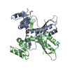
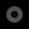
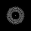
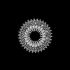
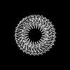
Journal: Commun Biol / Year: 2025 Title: Structural basis for assembly and function of the Salmonella flagellar MS-ring with three different symmetries. Authors: Miki Kinoshita / Fumiaki Makino / Tomoko Miyata / Katsumi Imada / Keiichi Namba / Tohru Minamino / Abstract: The flagellar MS-ring is the initial template for flagellar assembly and houses the flagellar protein export complex. The MS-ring has three parts of different symmetries within the ring structure by ...The flagellar MS-ring is the initial template for flagellar assembly and houses the flagellar protein export complex. The MS-ring has three parts of different symmetries within the ring structure by assembly of FliF subunits in two different conformations with distinct arrangements of three ring-building motifs, RBM1, RBM2, and RBM3. However, it remains unknown how these symmetries are generated. A combination of cryoEM structure and structure-based mutational analyses demonstrates that the well-conserved DQxGxxL motif in the RBM2-RBM3 hinge loop allows RBM2 to take two different orientations relative to RBM3. Of 34 FliF subunits of the MS-ring in the basal body, 23 RBM2 domains form an inner ring with a central pore that accommodates the flagellar protein export complex, and the remaining 11 RBM2 domains form 11 cog-like structures together with RBM1 domains just outside the inner RBM2-ring. We propose that a dimer of FliF with two different conformations initiates MS-ring assembly.
In the structure databanks used in Yorodumi, some data are registered as the other names, "COVID-19 virus" and "2019-nCoV". Here are the details of the virus and the list of structure data.
Jan 31, 2019. EMDB accession codes are about to change! (news from PDBe EMDB page)
EMDB accession codes are about to change! (news from PDBe EMDB page)
The allocation of 4 digits for EMDB accession codes will soon come to an end. Whilst these codes will remain in use, new EMDB accession codes will include an additional digit and will expand incrementally as the available range of codes is exhausted. The current 4-digit format prefixed with “EMD-” (i.e. EMD-XXXX) will advance to a 5-digit format (i.e. EMD-XXXXX), and so on. It is currently estimated that the 4-digit codes will be depleted around Spring 2019, at which point the 5-digit format will come into force.
The EM Navigator/Yorodumi systems omit the EMD- prefix.
Related info.:Q: What is EMD? / ID/Accession-code notation in Yorodumi/EM Navigator
Yorodumi is a browser for structure data from EMDB, PDB, SASBDB, etc.
This page is also the successor to EM Navigator detail page, and also detail information page/front-end page for Omokage search.
The word "yorodu" (or yorozu) is an old Japanese word meaning "ten thousand". "mi" (miru) is to see.
Related info.:EMDB / PDB / SASBDB / Comparison of 3 databanks / Yorodumi Search / Aug 31, 2016. New EM Navigator & Yorodumi / Yorodumi Papers / Jmol/JSmol / Function and homology information / Changes in new EM Navigator and Yorodumi
 Movie
Movie Controller

 Yorodumi
 Open data
 Basic information
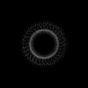
 Map data
 Sample
 Keywords
 Function and homology information
 Salmonella enterica subsp. enterica serovar Typhimurium str. LT2 (bacteria) /
  Authors
 Japan, 10 items
  Citation
 Structure visualization
 Downloads & links
 emd_60007.png
 http://ftp.pdbj.org/pub/emdb/structures/EMD-60007
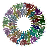


 Z (Sec.)
 Y (Row.)
 X (Col.)


 Sample components
 Processing
 Electron microscopy
 FIELD EMISSION GUN