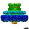
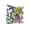
Journal: mBio / Year: 2021 Title: Two Distinct Conformations in 34 FliF Subunits Generate Three Different Symmetries within the Flagellar MS-Ring. Authors: Norihiro Takekawa / Akihiro Kawamoto / Mayuko Sakuma / Takayuki Kato / Seiji Kojima / Miki Kinoshita / Tohru Minamino / Keiichi Namba / Michio Homma / Katsumi Imada / Abstract: The bacterial flagellum is a protein nanomachine essential for bacterial motility. The flagellar basal body contains several ring structures. The MS-ring is embedded in the cytoplasmic membrane and ...The bacterial flagellum is a protein nanomachine essential for bacterial motility. The flagellar basal body contains several ring structures. The MS-ring is embedded in the cytoplasmic membrane and is formed at the earliest stage of flagellar formation to serve as the base for flagellar assembly as well as a housing for the flagellar protein export gate complex. The MS-ring is formed by FliF, which has two transmembrane helices and a large periplasmic region. A recent electron cryomicroscopy (cryoEM) study of the MS-ring formed by overexpressed FliF revealed a symmetry mismatch between the S-ring and inner part of the M-ring. However, the actual symmetry relation in the native MS-ring and positions of missing domains remain obscure. Here, we show the structure of the M-ring by combining cryoEM and X-ray crystallography. The crystal structure of the N-terminal half of the periplasmic region of FliF showed that it consists of two domains (D1 and D2) resembling PrgK D1/PrgH D2 and PrgK D2/PrgH D3 of the injectisome. CryoEM analysis revealed that the inner part of the M-ring shows a gear wheel-like density with the inner ring of C23 symmetry surrounded by cogs with C11 symmetry, to which 34 copies of FliF fitted well. We propose that FliF adopts two distinct orientations in the M-ring relative to the rest of FliF, with 23 chains forming the wheel and 11 chains forming the cogs, and the 34 chains come together to form the S-ring with C34 symmetry for multiple functions of the MS-ring. The bacterial flagellum is a motility organelle formed by tens of thousands of protein molecules. At the earliest stage of flagellar assembly, a transmembrane protein, FliF, forms the MS-ring in the cytoplasmic membrane as the base for flagellar assembly. Here, we solved the crystal structure of a FliF fragment. Electron cryomicroscopy (cryoEM) structural analysis of the MS-ring showed that the M-ring and S-ring have different rotational symmetries. By docking the crystal structure of the FliF fragment into the cryoEM density map of the entire MS-ring, we built a model of the whole periplasmic region of FliF and proposed that FliF adopts two distinct conformations to generate three distinct C11, C23, and C34 symmetries within the MS-ring for its multiple functions.
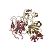
34 subunits of this protein form a single biological assembly. However, this protein adopts two different types of conformations that differ from the deposited crystal structure in the assembly. 23 subunits with one conformation form a ring, and 11 subunits with the other conformation surround the ring in the assembly. Therefore, the biological assembly cannot be described using a simple rotation and translation matrix.
-
Components
#1: Protein
FlagellarM-ringprotein / FliF
Mass: 19568.713 Da / Num. of mol.: 2 / Fragment: UNP residues 58-213 Source method: isolated from a genetically manipulated source Source: (gene. exp.) Aquifex aeolicus (strain VF5) (bacteria) Gene: fliF, aq_1182 / Plasmid: pCold I / Production host: Escherichia coli BL21(DE3) (bacteria) / References: UniProt: O67241
In the structure databanks used in Yorodumi, some data are registered as the other names, "COVID-19 virus" and "2019-nCoV". Here are the details of the virus and the list of structure data.
Jan 31, 2019. EMDB accession codes are about to change! (news from PDBe EMDB page)
EMDB accession codes are about to change! (news from PDBe EMDB page)
The allocation of 4 digits for EMDB accession codes will soon come to an end. Whilst these codes will remain in use, new EMDB accession codes will include an additional digit and will expand incrementally as the available range of codes is exhausted. The current 4-digit format prefixed with “EMD-” (i.e. EMD-XXXX) will advance to a 5-digit format (i.e. EMD-XXXXX), and so on. It is currently estimated that the 4-digit codes will be depleted around Spring 2019, at which point the 5-digit format will come into force.
The EM Navigator/Yorodumi systems omit the EMD- prefix.
Related info.:Q: What is EMD? / ID/Accession-code notation in Yorodumi/EM Navigator
Yorodumi is a browser for structure data from EMDB, PDB, SASBDB, etc.
This page is also the successor to EM Navigator detail page, and also detail information page/front-end page for Omokage search.
The word "yorodu" (or yorozu) is an old Japanese word meaning "ten thousand". "mi" (miru) is to see.
Related info.:EMDB / PDB / SASBDB / Comparison of 3 databanks / Yorodumi Search / Aug 31, 2016. New EM Navigator & Yorodumi / Yorodumi Papers / Jmol/JSmol / Function and homology information / Changes in new EM Navigator and Yorodumi
 Movie
Movie Controller


 Open data
 Basic information
 Components
 Keywords
 Function and homology information
 Aquifex aeolicus (bacteria)
 X-RAY DIFFRACTION /
  Authors
 Japan, 2items
  Citation
 Structure visualization
 Downloads & links
 Other downloads


 PDBj
PDBj
 Assembly

 Mass: 118.174 Da / Num. of mol.: 1 / Source method: obtained synthetically / Formula: C6H14O2 / Comment: precipitant*YM
 Mass: 18.015 Da / Num. of mol.: 156 / Source method: isolated from a natural source / Formula: H2O
 Sample preparation
 Processing