Movie
Movie Controller

[English] 日本語
 Yorodumi
- PDB-8yee: Cryo-EM structure of in vitro reconstituted Light-harvesting comp... -
+ Open data

- Basic information
| Entry | Database: PDB / ID: 8yee | ||||||||||||||||||
|---|---|---|---|---|---|---|---|---|---|---|---|---|---|---|---|---|---|---|---|
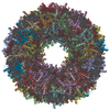
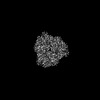
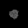
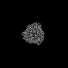
| Title | Cryo-EM structure of in vitro reconstituted Light-harvesting complex II trimer | ||||||||||||||||||
 Components Components | Chlorophyll a-b binding protein, chloroplastic | ||||||||||||||||||
 Keywords Keywords | PHOTOSYNTHESIS / Complex / light-harvesting / membrane protein | ||||||||||||||||||
| Function / homology |  Function and homology information Function and homology informationphotosynthesis, light harvesting in photosystem I / photosystem I / photosystem II / chlorophyll binding / chloroplast thylakoid membrane / response to light stimulus / metal ion binding Similarity search - Function | ||||||||||||||||||
| Biological species |  Spinacia oleracea (spinach) Spinacia oleracea (spinach) | ||||||||||||||||||
| Method | ELECTRON MICROSCOPY / single particle reconstruction / cryo EM / Resolution: 2.37 Å | ||||||||||||||||||
 Authors Authors | Seki, S. / Miyata, T. / Tanaka, H. / Namba, K. / Kurisu, G. / Fujii, R. | ||||||||||||||||||
| Funding support |  Japan, 5items Japan, 5items
| ||||||||||||||||||
 Citation Citation | Journal: PNAS Nexus / Year: 2024 Title: Structure-based validation of recombinant light-harvesting complex II. Authors: Soichiro Seki / Tomoko Miyata / Naoko Norioka / Hideaki Tanaka / Genji Kurisu / Keiichi Namba / Ritsuko Fujii / Abstract: Light-harvesting complex II (LHCII) captures sunlight and dissipates excess energy to drive photosynthesis. To elucidate this mechanism, the individual optical properties of pigments in the LHCII ...Light-harvesting complex II (LHCII) captures sunlight and dissipates excess energy to drive photosynthesis. To elucidate this mechanism, the individual optical properties of pigments in the LHCII protein must be identified. In vitro reconstitution with apoproteins synthesized by and pigment-lipid mixtures from natural sources is an effective approach; however, the local environment surrounding each pigment within reconstituted LHCII (rLHCII) has only been indirectly estimated using spectroscopic and biochemical methods. Here, we used cryo-electron microscopy to determine the 3D structure of the rLHCII trimer and found that rLHCII exhibited a structure that was virtually identical to that of native LHCII, with a few exceptions: some C-terminal amino acids were not visible, likely due to aggregation of the His-tags; a carotenoid at the V1 site was not visible; and at site 614 showed mixed occupancy by both chlorophyll and molecules. Our observations confirmed the applicability of the in vitro reconstitution technique. | ||||||||||||||||||
| History |
|
- Structure visualization
| Structure viewer | Molecule: MolmilJmol/JSmol |
|---|
- Downloads & links
-Download
| PDBx/mmCIF format | 8yee.cif.gz | 191.2 KB | Display | PDBx/mmCIF format |
|---|---|---|---|---|
| PDB format | pdb8yee.ent.gz | Display | PDB format | |
| PDBx/mmJSON format | 8yee.json.gz | Tree view | PDBx/mmJSON format | |
| Others |  Other downloads Other downloads |
-Validation report
| Arichive directory | https://data.pdbj.org/pub/pdb/validation_reports/ye/8yeeftp://data.pdbj.org/pub/pdb/validation_reports/ye/8yee | HTTPS FTP |
|---|
-Related structure data
| Related structure data | 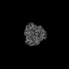 39192MC 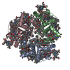 8y15C M: map data used to model this data C: citing same article ( |
|---|---|
| Similar structure data |
-Links
 PDBj
PDBj


- Assembly
| Deposited unit | 
|
|---|---|
| 1 |
|
-Components
-Protein , 1 types, 3 molecules ABC
| #1: Protein | Mass: 24455.611 Da / Num. of mol.: 3 Source method: isolated from a genetically manipulated source Source: (gene. exp.) Spinacia oleracea (spinach) / Production host:  |
|---|
-Non-polymers , 6 types, 180 molecules 


| #2: Chemical | ChemComp-CHL /  Mass: 907.472 Da / Num. of mol.: 18 / Source method: obtained synthetically / Formula: C55H70MgN4O6 / Feature type: SUBJECT OF INVESTIGATION Mass: 907.472 Da / Num. of mol.: 18 / Source method: obtained synthetically / Formula: C55H70MgN4O6 / Feature type: SUBJECT OF INVESTIGATION#3: Chemical | ChemComp-CLA /  Mass: 893.489 Da / Num. of mol.: 24 / Source method: obtained synthetically / Formula: C55H72MgN4O5 / Feature type: SUBJECT OF INVESTIGATION Mass: 893.489 Da / Num. of mol.: 24 / Source method: obtained synthetically / Formula: C55H72MgN4O5 / Feature type: SUBJECT OF INVESTIGATION#4: Chemical | ChemComp-A1LXP / Mass: 568.871 Da / Num. of mol.: 6 / Source method: obtained synthetically / Formula: C40H56O2 / Feature type: SUBJECT OF INVESTIGATION #5: Chemical |  Mass: 600.870 Da / Num. of mol.: 3 / Source method: obtained synthetically / Formula: C40H56O4 Mass: 600.870 Da / Num. of mol.: 3 / Source method: obtained synthetically / Formula: C40H56O4#6: Chemical |  Mass: 722.970 Da / Num. of mol.: 3 / Source method: obtained synthetically / Formula: C38H75O10P / Comment: phospholipid*YM Mass: 722.970 Da / Num. of mol.: 3 / Source method: obtained synthetically / Formula: C38H75O10P / Comment: phospholipid*YM#7: Water | ChemComp-HOH / | Mass: 18.015 Da / Num. of mol.: 126 / Source method: isolated from a natural source / Formula: H2O |
|---|
-Details
| Has ligand of interest | Y |
|---|---|
| Has protein modification | N |
-Experimental details
-Experiment
| Experiment | Method: ELECTRON MICROSCOPY |
|---|---|
| EM experiment | Aggregation state: PARTICLE / 3D reconstruction method: single particle reconstruction |
- Sample preparation
| Component | Name: In vitro reconstituted Light-harvesting complex II / Type: COMPLEX / Entity ID: #1 / Source: RECOMBINANT |
|---|---|
| Molecular weight | Value: 0.12 MDa / Experimental value: NO |
| Source (natural) | Organism: Spinacia oleracea (spinach) |
| Source (recombinant) | Organism: |
| Buffer solution | pH: 7.5 / Details: 0.03 % n-Dodexyl-alpha-D-maltoside |
| Buffer component | Conc.: 25 mM / Name: HEPES / Formula: C8H18N2O4S |
| Specimen | Conc.: 5 mg/ml / Embedding applied: NO / Shadowing applied: NO / Staining applied: NO / Vitrification applied: YES |
| Specimen support | Grid material: COPPER / Grid mesh size: 200 divisions/in. / Grid type: Quantifoil R1.2/1.3 |
| Vitrification | Instrument: FEI VITROBOT MARK IV / Cryogen name: ETHANE / Humidity: 100 % / Chamber temperature: 277 K |
- Electron microscopy imaging
| Microscopy | Model: JEOL CRYO ARM 300 |
|---|---|
| Electron gun | Electron source:  FIELD EMISSION GUN / Accelerating voltage: 300 kV / Illumination mode: FLOOD BEAM FIELD EMISSION GUN / Accelerating voltage: 300 kV / Illumination mode: FLOOD BEAM |
| Electron lens | Mode: BRIGHT FIELD / Nominal magnification: 60000 X / Nominal defocus max: 2200 nm / Nominal defocus min: 700 nm / Cs: 2.7 mm |
| Specimen holder | Cryogen: NITROGEN / Specimen holder model: JEOL CRYOSPECPORTER |
| Image recording | Average exposure time: 3 sec. / Electron dose: 80 e/Å2 / Film or detector model: GATAN K3 (6k x 4k) / Num. of grids imaged: 1 / Num. of real images: 6145 |
| EM imaging optics | Energyfilter name: In-column Omega Filter / Energyfilter slit width: 20 eV |
- Processing
| EM software |
| ||||||||||||||||||||||||||||||||||||||||
|---|---|---|---|---|---|---|---|---|---|---|---|---|---|---|---|---|---|---|---|---|---|---|---|---|---|---|---|---|---|---|---|---|---|---|---|---|---|---|---|---|---|
| CTF correction | Type: PHASE FLIPPING AND AMPLITUDE CORRECTION | ||||||||||||||||||||||||||||||||||||||||
| Particle selection | Num. of particles selected: 4734387 | ||||||||||||||||||||||||||||||||||||||||
| Symmetry | Point symmetry: C3 (3 fold cyclic) | ||||||||||||||||||||||||||||||||||||||||
| 3D reconstruction | Resolution: 2.37 Å / Resolution method: FSC 0.143 CUT-OFF / Num. of particles: 179318 / Algorithm: FOURIER SPACE / Symmetry type: POINT | ||||||||||||||||||||||||||||||||||||||||
| Atomic model building | Space: REAL | ||||||||||||||||||||||||||||||||||||||||
| Atomic model building | PDB-ID: 1RWT Pdb chain-ID: B / Accession code: 1RWT / Chain residue range: 14-229 / Pdb chain residue range: 14-229 / Source name: PDB / Type: experimental model | ||||||||||||||||||||||||||||||||||||||||
| Refine LS restraints |
|