+ データを開く
- 基本情報
基本情報
| 登録情報 | データベース: PDB / ID: 8uxg | ||||||
|---|---|---|---|---|---|---|---|
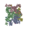
| タイトル | Structure of PKA phosphorylated human RyR2-R420W in the closed state in the presence of ARM210 | ||||||
 要素 要素 |
| ||||||
 キーワード キーワード | TRANSPORT PROTEIN / calcium channel | ||||||
| 機能・相同性 |  機能・相同性情報 機能・相同性情報junctional sarcoplasmic reticulum membrane / sarcoplasmic reticulum calcium ion transport / establishment of protein localization to endoplasmic reticulum / type B pancreatic cell apoptotic process / Purkinje myocyte to ventricular cardiac muscle cell signaling / calcium-induced calcium release activity / regulation of atrial cardiac muscle cell action potential / left ventricular cardiac muscle tissue morphogenesis / suramin binding / regulation of AV node cell action potential ...junctional sarcoplasmic reticulum membrane / sarcoplasmic reticulum calcium ion transport / establishment of protein localization to endoplasmic reticulum / type B pancreatic cell apoptotic process / Purkinje myocyte to ventricular cardiac muscle cell signaling / calcium-induced calcium release activity / regulation of atrial cardiac muscle cell action potential / left ventricular cardiac muscle tissue morphogenesis / suramin binding / regulation of AV node cell action potential / regulation of SA node cell action potential / cell communication by electrical coupling involved in cardiac conduction / regulation of ventricular cardiac muscle cell action potential / ventricular cardiac muscle cell action potential / positive regulation of sequestering of calcium ion / negative regulation of calcium-mediated signaling / embryonic heart tube morphogenesis / cardiac muscle hypertrophy / negative regulation of insulin secretion involved in cellular response to glucose stimulus / neuronal action potential propagation / negative regulation of release of sequestered calcium ion into cytosol / insulin secretion involved in cellular response to glucose stimulus / calcium ion transport into cytosol / ryanodine-sensitive calcium-release channel activity / response to caffeine / regulation of cardiac muscle contraction by calcium ion signaling / release of sequestered calcium ion into cytosol by sarcoplasmic reticulum / response to redox state / 'de novo' protein folding / cellular response to caffeine / negative regulation of heart rate / FK506 binding / response to muscle activity / protein kinase A regulatory subunit binding / protein kinase A catalytic subunit binding / positive regulation of the force of heart contraction / smooth endoplasmic reticulum / intracellularly gated calcium channel activity / smooth muscle contraction / detection of calcium ion / regulation of cardiac muscle contraction / regulation of cytosolic calcium ion concentration / T cell proliferation / regulation of cardiac muscle contraction by regulation of the release of sequestered calcium ion / positive regulation of heart rate / calcium channel inhibitor activity / Ion homeostasis / cardiac muscle contraction / regulation of release of sequestered calcium ion into cytosol by sarcoplasmic reticulum / response to muscle stretch / release of sequestered calcium ion into cytosol / cellular response to epinephrine stimulus / calcium channel complex / sarcoplasmic reticulum membrane / regulation of heart rate / sarcoplasmic reticulum / protein maturation / peptidylprolyl isomerase / calcium channel regulator activity / peptidyl-prolyl cis-trans isomerase activity / establishment of localization in cell / Stimuli-sensing channels / calcium-mediated signaling / sarcolemma / calcium channel activity / Z disc / intracellular calcium ion homeostasis / calcium ion transport / positive regulation of cytosolic calcium ion concentration / protein refolding / transmembrane transporter binding / calmodulin binding / response to hypoxia / signaling receptor binding / calcium ion binding / enzyme binding / protein-containing complex / identical protein binding / membrane / plasma membrane / cytoplasm 類似検索 - 分子機能 | ||||||
| 生物種 |  Homo sapiens (ヒト) Homo sapiens (ヒト) | ||||||
| 手法 | 電子顕微鏡法 / 単粒子再構成法 / クライオ電子顕微鏡法 / 解像度: 3.08 Å | ||||||
 データ登録者 データ登録者 | Miotto, M.C. / Marks, A.R. | ||||||
| 資金援助 |  米国, 1件 米国, 1件
| ||||||
 引用 引用 |  ジャーナル: Nat Commun / 年: 2024 ジャーナル: Nat Commun / 年: 2024タイトル: Structural basis for ryanodine receptor type 2 leak in heart failure and arrhythmogenic disorders. 著者: Marco C Miotto / Steven Reiken / Anetta Wronska / Qi Yuan / Haikel Dridi / Yang Liu / Gunnar Weninger / Carl Tchagou / Andrew R Marks /  要旨: Heart failure, the leading cause of mortality and morbidity in the developed world, is characterized by cardiac ryanodine receptor 2 channels that are hyperphosphorylated, oxidized, and depleted of ...Heart failure, the leading cause of mortality and morbidity in the developed world, is characterized by cardiac ryanodine receptor 2 channels that are hyperphosphorylated, oxidized, and depleted of the stabilizing subunit calstabin-2. This results in a diastolic sarcoplasmic reticulum Ca leak that impairs cardiac contractility and triggers arrhythmias. Genetic mutations in ryanodine receptor 2 can also cause Ca leak, leading to arrhythmias and sudden cardiac death. Here, we solved the cryogenic electron microscopy structures of ryanodine receptor 2 variants linked either to heart failure or inherited sudden cardiac death. All are in the primed state, part way between closed and open. Binding of Rycal drugs to ryanodine receptor 2 channels reverts the primed state back towards the closed state, decreasing Ca leak, improving cardiac function, and preventing arrhythmias. We propose a structural-physiological mechanism whereby the ryanodine receptor 2 channel primed state underlies the arrhythmias in heart failure and arrhythmogenic disorders. | ||||||
| 履歴 |
|
- 構造の表示
構造の表示
| 構造ビューア | 分子:  Molmil Molmil Jmol/JSmol Jmol/JSmol |
|---|
- ダウンロードとリンク
- ダウンロード
| PDBx/mmCIF形式 |  8uxg.cif.gz 8uxg.cif.gz | 2.9 MB | 表示 |  PDBx/mmCIF形式 PDBx/mmCIF形式 |
|---|---|---|---|---|
| PDB形式 |  pdb8uxg.ent.gz pdb8uxg.ent.gz | 表示 |  PDB形式 PDB形式 | |
| PDBx/mmJSON形式 |  8uxg.json.gz 8uxg.json.gz | ツリー表示 |  PDBx/mmJSON形式 PDBx/mmJSON形式 | |
| その他 |  その他のダウンロード その他のダウンロード |
-検証レポート
| 文書・要旨 |  8uxg_validation.pdf.gz 8uxg_validation.pdf.gz | 1.6 MB | 表示 |  wwPDB検証レポート wwPDB検証レポート |
|---|---|---|---|---|
| 文書・詳細版 |  8uxg_full_validation.pdf.gz 8uxg_full_validation.pdf.gz | 1.9 MB | 表示 | |
| XML形式データ |  8uxg_validation.xml.gz 8uxg_validation.xml.gz | 406 KB | 表示 | |
| CIF形式データ |  8uxg_validation.cif.gz 8uxg_validation.cif.gz | 625.5 KB | 表示 | |
| アーカイブディレクトリ |  https://data.pdbj.org/pub/pdb/validation_reports/ux/8uxg https://data.pdbj.org/pub/pdb/validation_reports/ux/8uxg ftp://data.pdbj.org/pub/pdb/validation_reports/ux/8uxg ftp://data.pdbj.org/pub/pdb/validation_reports/ux/8uxg | HTTPS FTP |
-関連構造データ
| 関連構造データ |  42763MC  8uq2C  8uq3C  8uq4C  8uq5C  8uxcC  8uxeC  8uxfC  8uxhC  8uxiC  8uxlC  8uxmC C: 同じ文献を引用 ( M: このデータのモデリングに利用したマップデータ |
|---|---|
| 類似構造データ | 類似検索 - 機能・相同性  F&H 検索 F&H 検索 |
- リンク
リンク
- 集合体
集合体
| 登録構造単位 | 
|
|---|---|
| 1 |
|
- 要素
要素
| #1: タンパク質 | 分子量: 565315.125 Da / 分子数: 4 / 由来タイプ: 組換発現 / 由来: (組換発現)  Homo sapiens (ヒト) / 遺伝子: RYR2 / 細胞株 (発現宿主): HEK293 / 発現宿主: Homo sapiens (ヒト) / 遺伝子: RYR2 / 細胞株 (発現宿主): HEK293 / 発現宿主:  Homo sapiens (ヒト) / 参照: UniProt: Q92736 Homo sapiens (ヒト) / 参照: UniProt: Q92736#2: タンパク質 | 分子量: 11798.501 Da / 分子数: 4 / 由来タイプ: 組換発現 / 由来: (組換発現)  Homo sapiens (ヒト) / 遺伝子: FKBP1B, FKBP12.6, FKBP1L, FKBP9, OTK4 / 発現宿主: Homo sapiens (ヒト) / 遺伝子: FKBP1B, FKBP12.6, FKBP1L, FKBP9, OTK4 / 発現宿主:  #3: 化合物 | ChemComp-ZN / #4: 化合物 | ChemComp-ATP / #5: 化合物 | ChemComp-KVR / 研究の焦点であるリガンドがあるか | Y | Has protein modification | Y | |
|---|
-実験情報
-実験
| 実験 | 手法: 電子顕微鏡法 |
|---|---|
| EM実験 | 試料の集合状態: PARTICLE / 3次元再構成法: 単粒子再構成法 |
- 試料調製
試料調製
| 構成要素 |
| |||||||||||||||||||||||||||||||||||||||||||||
|---|---|---|---|---|---|---|---|---|---|---|---|---|---|---|---|---|---|---|---|---|---|---|---|---|---|---|---|---|---|---|---|---|---|---|---|---|---|---|---|---|---|---|---|---|---|---|
| 由来(天然) |
| |||||||||||||||||||||||||||||||||||||||||||||
| 由来(組換発現) |
| |||||||||||||||||||||||||||||||||||||||||||||
| 緩衝液 | pH: 7.4 | |||||||||||||||||||||||||||||||||||||||||||||
| 緩衝液成分 |
| |||||||||||||||||||||||||||||||||||||||||||||
| 試料 | 濃度: 2.5 mg/ml / 包埋: NO / シャドウイング: NO / 染色: NO / 凍結: YES | |||||||||||||||||||||||||||||||||||||||||||||
| 急速凍結 | 凍結剤: ETHANE |
- 電子顕微鏡撮影
| 実験機器 |  モデル: Titan Krios / 画像提供: FEI Company |
|---|---|
| 顕微鏡 | モデル: FEI TITAN KRIOS |
| 電子銃 | 電子線源:  FIELD EMISSION GUN / 加速電圧: 300 kV / 照射モード: FLOOD BEAM FIELD EMISSION GUN / 加速電圧: 300 kV / 照射モード: FLOOD BEAM |
| 電子レンズ | モード: BRIGHT FIELD / 最大 デフォーカス(公称値): 1200 nm / 最小 デフォーカス(公称値): 500 nm / Cs: 2.7 mm / C2レンズ絞り径: 100 µm |
| 試料ホルダ | 凍結剤: NITROGEN 試料ホルダーモデル: FEI TITAN KRIOS AUTOGRID HOLDER 最高温度: 100 K / 最低温度: 80 K |
| 撮影 | 電子線照射量: 58 e/Å2 フィルム・検出器のモデル: GATAN K3 BIOQUANTUM (6k x 4k) |
| 電子光学装置 | エネルギーフィルター名称: GIF Bioquantum / エネルギーフィルタースリット幅: 20 eV |
| 画像スキャン | サンプリングサイズ: 5 µm / 横: 5760 / 縦: 4092 |
- 解析
解析
| EMソフトウェア |
| |||||||||||||||||||||||||||||||||||||||||||||
|---|---|---|---|---|---|---|---|---|---|---|---|---|---|---|---|---|---|---|---|---|---|---|---|---|---|---|---|---|---|---|---|---|---|---|---|---|---|---|---|---|---|---|---|---|---|---|
| CTF補正 | タイプ: PHASE FLIPPING AND AMPLITUDE CORRECTION | |||||||||||||||||||||||||||||||||||||||||||||
| 対称性 | 点対称性: C4 (4回回転対称) | |||||||||||||||||||||||||||||||||||||||||||||
| 3次元再構成 | 解像度: 3.08 Å / 解像度の算出法: FSC 0.143 CUT-OFF / 粒子像の数: 181724 / 対称性のタイプ: POINT | |||||||||||||||||||||||||||||||||||||||||||||
| 原子モデル構築 | PDB-ID: 7UA5 Accession code: 7UA5 / Source name: PDB / タイプ: experimental model | |||||||||||||||||||||||||||||||||||||||||||||
| 拘束条件 |
|
 ムービー
ムービー コントローラー


 PDBj
PDBj


