 Movie
Movie Controller


+ Open data

- Basic information
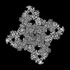
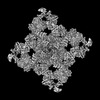
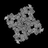
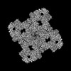
| Entry | Database: PDB / ID: 7ua5 | ||||||
|---|---|---|---|---|---|---|---|
| Title | Structure of dephosphorylated human RyR2 in the closed state | ||||||
 Components Components |
| ||||||
 Keywords Keywords | MEMBRANE PROTEIN / calcium channel | ||||||
| Function / homology |  Function and homology information Function and homology informationjunctional sarcoplasmic reticulum membrane / type B pancreatic cell apoptotic process / Purkinje myocyte to ventricular cardiac muscle cell signaling / regulation of atrial cardiac muscle cell action potential / left ventricular cardiac muscle tissue morphogenesis / suramin binding / regulation of AV node cell action potential / sarcoplasmic reticulum calcium ion transport / regulation of SA node cell action potential / calcium-induced calcium release activity ...junctional sarcoplasmic reticulum membrane / type B pancreatic cell apoptotic process / Purkinje myocyte to ventricular cardiac muscle cell signaling / regulation of atrial cardiac muscle cell action potential / left ventricular cardiac muscle tissue morphogenesis / suramin binding / regulation of AV node cell action potential / sarcoplasmic reticulum calcium ion transport / regulation of SA node cell action potential / calcium-induced calcium release activity / calcium ion transport into cytosol / embryonic heart tube morphogenesis / cell communication by electrical coupling involved in cardiac conduction / ventricular cardiac muscle cell action potential / negative regulation of calcium-mediated signaling / cardiac muscle hypertrophy / regulation of ventricular cardiac muscle cell action potential / ryanodine-sensitive calcium-release channel activity / release of sequestered calcium ion into cytosol by sarcoplasmic reticulum / negative regulation of release of sequestered calcium ion into cytosol / response to redox state / regulation of cardiac muscle contraction by calcium ion signaling / negative regulation of heart rate / cellular response to caffeine / response to caffeine / response to muscle activity / 'de novo' protein folding / FK506 binding / protein kinase A regulatory subunit binding / protein kinase A catalytic subunit binding / positive regulation of the force of heart contraction / smooth endoplasmic reticulum / intracellularly gated calcium channel activity / regulation of ryanodine-sensitive calcium-release channel activity / detection of calcium ion / regulation of cardiac muscle contraction / response to muscle stretch / regulation of cytosolic calcium ion concentration / positive regulation of heart rate / cardiac muscle contraction / release of sequestered calcium ion into cytosol / calcium channel inhibitor activity / regulation of release of sequestered calcium ion into cytosol by sarcoplasmic reticulum / Ion homeostasis / regulation of cardiac muscle contraction by regulation of the release of sequestered calcium ion / cellular response to epinephrine stimulus / sarcoplasmic reticulum membrane / calcium channel complex / regulation of heart rate / calcium-mediated signaling / peptidylprolyl isomerase / sarcoplasmic reticulum / striated muscle contraction / peptidyl-prolyl cis-trans isomerase activity / calcium channel regulator activity / protein maturation / sarcolemma / intracellular calcium ion homeostasis / Stimuli-sensing channels / Z disc / calcium channel activity / calcium ion transport / protein refolding / protein folding / response to hypoxia / transmembrane transporter binding / calmodulin binding / signaling receptor binding / calcium ion binding / enzyme binding / protein-containing complex / membrane / identical protein binding / plasma membrane / cytoplasm Similarity search - Function | ||||||
| Biological species |  Homo sapiens (human) Homo sapiens (human) | ||||||
| Method | ELECTRON MICROSCOPY / single particle reconstruction / cryo EM / Resolution: 2.83 Å | ||||||
 Authors Authors | Miotto, M.C. / Marks, A.R. | ||||||
| Funding support |  United States, 1items United States, 1items
| ||||||
 Citation Citation | Journal: Sci Adv / Year: 2022 Title: Structural analyses of human ryanodine receptor type 2 channels reveal the mechanisms for sudden cardiac death and treatment. Authors: Marco C Miotto / Gunnar Weninger / Haikel Dridi / Qi Yuan / Yang Liu / Anetta Wronska / Zephan Melville / Leah Sittenfeld / Steven Reiken / Andrew R Marks / Abstract: Ryanodine receptor type 2 (RyR2) mutations have been linked to an inherited form of exercise-induced sudden cardiac death called catecholaminergic polymorphic ventricular tachycardia (CPVT). CPVT ...Ryanodine receptor type 2 (RyR2) mutations have been linked to an inherited form of exercise-induced sudden cardiac death called catecholaminergic polymorphic ventricular tachycardia (CPVT). CPVT results from stress-induced sarcoplasmic reticular Ca leak via the mutant RyR2 channels during diastole. We present atomic models of human wild-type (WT) RyR2 and the CPVT mutant RyR2-R2474S determined by cryo-electron microscopy with overall resolutions in the range of 2.6 to 3.6 Å, and reaching local resolutions of 2.25 Å, unprecedented for RyR2 channels. Under nonactivating conditions, the RyR2-R2474S channel is in a "primed" state between the closed and open states of WT RyR2, rendering it more sensitive to activation that results in stress-induced Ca leak. The Rycal drug ARM210 binds to RyR2-R2474S, reverting the primed state toward the closed state. Together, these studies provide a mechanism for CPVT and for the therapeutic actions of ARM210. | ||||||
| History |
|
- Structure visualization
| Structure viewer | Molecule: MolmilJmol/JSmol |
|---|
- Downloads & links
-Download
| PDBx/mmCIF format | 7ua5.cif.gz | 3 MB | Display | PDBx/mmCIF format |
|---|---|---|---|---|
| PDB format | pdb7ua5.ent.gz | Display | PDB format | |
| PDBx/mmJSON format | 7ua5.json.gz | Tree view | PDBx/mmJSON format | |
| Others |  Other downloads Other downloads |
-Validation report
| Arichive directory | https://data.pdbj.org/pub/pdb/validation_reports/ua/7ua5ftp://data.pdbj.org/pub/pdb/validation_reports/ua/7ua5 | HTTPS FTP |
|---|
-Related structure data
| Related structure data |  26415MC  7u9qC  7u9rC  7u9tC  7u9xC  7u9zC  7ua1C  7ua3C  7ua4C  7ua9C M: map data used to model this data C: citing same article ( |
|---|---|
| Similar structure data |


-Links
 PDBj
PDBj


- Assembly
| Deposited unit | 
| |||||||||||||||||||||||||||||||||||||||||||||||||||||||||||||||||||||||||||||||||||||||||||||||||||||||||||||||||||||||||||||||||
|---|---|---|---|---|---|---|---|---|---|---|---|---|---|---|---|---|---|---|---|---|---|---|---|---|---|---|---|---|---|---|---|---|---|---|---|---|---|---|---|---|---|---|---|---|---|---|---|---|---|---|---|---|---|---|---|---|---|---|---|---|---|---|---|---|---|---|---|---|---|---|---|---|---|---|---|---|---|---|---|---|---|---|---|---|---|---|---|---|---|---|---|---|---|---|---|---|---|---|---|---|---|---|---|---|---|---|---|---|---|---|---|---|---|---|---|---|---|---|---|---|---|---|---|---|---|---|---|---|---|---|
| 1 |
| |||||||||||||||||||||||||||||||||||||||||||||||||||||||||||||||||||||||||||||||||||||||||||||||||||||||||||||||||||||||||||||||||
| Noncrystallographic symmetry (NCS) | NCS domain:
NCS domain segments:
|