 Movie
Movie Controller

[English] 日本語
 Yorodumi
- PDB-7u9t: Structure of PKA phosphorylated human RyR2 in the closed state in... -
+ Open data

- Basic information
| Entry | Database: PDB / ID: 7u9t | ||||||
|---|---|---|---|---|---|---|---|
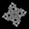
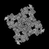
| Title | Structure of PKA phosphorylated human RyR2 in the closed state in the presence of Calmodulin | ||||||
 Components Components |
| ||||||
 Keywords Keywords | MEMBRANE PROTEIN / calcium channel | ||||||
| Function / homology |  Function and homology information Function and homology informationestablishment of protein localization to endoplasmic reticulum / junctional sarcoplasmic reticulum membrane / type B pancreatic cell apoptotic process / Purkinje myocyte to ventricular cardiac muscle cell signaling / regulation of atrial cardiac muscle cell action potential / left ventricular cardiac muscle tissue morphogenesis / suramin binding / regulation of AV node cell action potential / sarcoplasmic reticulum calcium ion transport / regulation of SA node cell action potential ...establishment of protein localization to endoplasmic reticulum / junctional sarcoplasmic reticulum membrane / type B pancreatic cell apoptotic process / Purkinje myocyte to ventricular cardiac muscle cell signaling / regulation of atrial cardiac muscle cell action potential / left ventricular cardiac muscle tissue morphogenesis / suramin binding / regulation of AV node cell action potential / sarcoplasmic reticulum calcium ion transport / regulation of SA node cell action potential / calcium-induced calcium release activity / cell communication by electrical coupling involved in cardiac conduction / : / ventricular cardiac muscle cell action potential / negative regulation of calcium-mediated signaling / regulation of ventricular cardiac muscle cell action potential / cardiac muscle hypertrophy / embryonic heart tube morphogenesis / negative regulation of insulin secretion involved in cellular response to glucose stimulus / calcium ion transport into cytosol / neuronal action potential propagation / insulin secretion involved in cellular response to glucose stimulus / ryanodine-sensitive calcium-release channel activity / release of sequestered calcium ion into cytosol by sarcoplasmic reticulum / negative regulation of release of sequestered calcium ion into cytosol / response to redox state / regulation of cardiac muscle contraction by calcium ion signaling / CaM pathway / Cam-PDE 1 activation / cellular response to caffeine / Sodium/Calcium exchangers / negative regulation of heart rate / Calmodulin induced events / 'de novo' protein folding / response to caffeine / Reduction of cytosolic Ca++ levels / Activation of Ca-permeable Kainate Receptor / CREB1 phosphorylation through the activation of CaMKII/CaMKK/CaMKIV cascasde / Loss of phosphorylation of MECP2 at T308 / CREB1 phosphorylation through the activation of Adenylate Cyclase / negative regulation of high voltage-gated calcium channel activity / PKA activation / CaMK IV-mediated phosphorylation of CREB / response to muscle activity / FK506 binding / Glycogen breakdown (glycogenolysis) / CLEC7A (Dectin-1) induces NFAT activation / negative regulation of ryanodine-sensitive calcium-release channel activity / organelle localization by membrane tethering / Activation of RAC1 downstream of NMDARs / : / autophagosome membrane docking / protein kinase A regulatory subunit binding / protein kinase A catalytic subunit binding / negative regulation of calcium ion export across plasma membrane / regulation of ryanodine-sensitive calcium-release channel activity / regulation of cardiac muscle cell action potential / positive regulation of the force of heart contraction / presynaptic endocytosis / Synthesis of IP3 and IP4 in the cytosol / intracellularly gated calcium channel activity / smooth endoplasmic reticulum / Phase 0 - rapid depolarisation / Negative regulation of NMDA receptor-mediated neuronal transmission / Unblocking of NMDA receptors, glutamate binding and activation / RHO GTPases activate PAKs / calcineurin-mediated signaling / regulation of cell communication by electrical coupling involved in cardiac conduction / smooth muscle contraction / Ion transport by P-type ATPases / Uptake and function of anthrax toxins / protein phosphatase activator activity / Long-term potentiation / Calcineurin activates NFAT / T cell proliferation / Regulation of MECP2 expression and activity / DARPP-32 events / Smooth Muscle Contraction / detection of calcium ion / regulation of cardiac muscle contraction / regulation of cytosolic calcium ion concentration / catalytic complex / positive regulation of heart rate / RHO GTPases activate IQGAPs / calcium channel inhibitor activity / presynaptic cytosol / striated muscle contraction / Activation of AMPK downstream of NMDARs / cellular response to interferon-beta / regulation of release of sequestered calcium ion into cytosol by sarcoplasmic reticulum / Ion homeostasis / response to muscle stretch / eNOS activation / cardiac muscle contraction / Tetrahydrobiopterin (BH4) synthesis, recycling, salvage and regulation / titin binding / Protein methylation / release of sequestered calcium ion into cytosol / regulation of cardiac muscle contraction by regulation of the release of sequestered calcium ion / regulation of calcium-mediated signaling Similarity search - Function | ||||||
| Biological species |  Homo sapiens (human) Homo sapiens (human) | ||||||
| Method | ELECTRON MICROSCOPY / single particle reconstruction / cryo EM / Resolution: 2.68 Å | ||||||
 Authors Authors | Miotto, M.C. / Marks, A.R. | ||||||
| Funding support |  United States, 1items United States, 1items
| ||||||
 Citation Citation | Journal: Sci Adv / Year: 2022 Title: Structural analyses of human ryanodine receptor type 2 channels reveal the mechanisms for sudden cardiac death and treatment. Authors: Marco C Miotto / Gunnar Weninger / Haikel Dridi / Qi Yuan / Yang Liu / Anetta Wronska / Zephan Melville / Leah Sittenfeld / Steven Reiken / Andrew R Marks / Abstract: Ryanodine receptor type 2 (RyR2) mutations have been linked to an inherited form of exercise-induced sudden cardiac death called catecholaminergic polymorphic ventricular tachycardia (CPVT). CPVT ...Ryanodine receptor type 2 (RyR2) mutations have been linked to an inherited form of exercise-induced sudden cardiac death called catecholaminergic polymorphic ventricular tachycardia (CPVT). CPVT results from stress-induced sarcoplasmic reticular Ca leak via the mutant RyR2 channels during diastole. We present atomic models of human wild-type (WT) RyR2 and the CPVT mutant RyR2-R2474S determined by cryo-electron microscopy with overall resolutions in the range of 2.6 to 3.6 Å, and reaching local resolutions of 2.25 Å, unprecedented for RyR2 channels. Under nonactivating conditions, the RyR2-R2474S channel is in a "primed" state between the closed and open states of WT RyR2, rendering it more sensitive to activation that results in stress-induced Ca leak. The Rycal drug ARM210 binds to RyR2-R2474S, reverting the primed state toward the closed state. Together, these studies provide a mechanism for CPVT and for the therapeutic actions of ARM210. | ||||||
| History |
|
- Structure visualization
| Structure viewer | Molecule: MolmilJmol/JSmol |
|---|
- Downloads & links
-Download
| PDBx/mmCIF format | 7u9t.cif.gz | 3 MB | Display | PDBx/mmCIF format |
|---|---|---|---|---|
| PDB format | pdb7u9t.ent.gz | Display | PDB format | |
| PDBx/mmJSON format | 7u9t.json.gz | Tree view | PDBx/mmJSON format | |
| Others |  Other downloads Other downloads |
-Validation report
| Arichive directory | https://data.pdbj.org/pub/pdb/validation_reports/u9/7u9tftp://data.pdbj.org/pub/pdb/validation_reports/u9/7u9t | HTTPS FTP |
|---|
-Related structure data
| Related structure data | 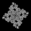 26408MC  7u9qC  7u9rC  7u9xC  7u9zC  7ua1C  7ua3C  7ua4C 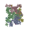 7ua5C  7ua9C M: map data used to model this data C: citing same article ( |
|---|---|
| Similar structure data |


-Links
 PDBj
PDBj


- Assembly
| Deposited unit | 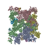
|
|---|---|
| 1 |
|
-Components
| #1: Protein | Mass: 11798.501 Da / Num. of mol.: 4 Source method: isolated from a genetically manipulated source Source: (gene. exp.) Homo sapiens (human) / Gene: FKBP1B, FKBP12.6, FKBP1L, FKBP9, OTK4 / Production host:  #2: Protein | Mass: 565286.125 Da / Num. of mol.: 4 Source method: isolated from a genetically manipulated source Source: (gene. exp.) Homo sapiens (human) / Gene: RYR2 / Cell line (production host): HEK293 / Production host: Homo sapiens (human) / References: UniProt: Q92736#3: Protein | Mass: 16852.545 Da / Num. of mol.: 4 Source method: isolated from a genetically manipulated source Source: (gene. exp.) Homo sapiens (human) / Gene: CALM1, CALM, CAM, CAM1 / Production host: #4: Chemical | ChemComp-ZN /   Mass: 65.409 Da / Num. of mol.: 4 / Source method: obtained synthetically / Formula: Zn / Feature type: SUBJECT OF INVESTIGATION Mass: 65.409 Da / Num. of mol.: 4 / Source method: obtained synthetically / Formula: Zn / Feature type: SUBJECT OF INVESTIGATION#5: Chemical | ChemComp-ATP /   Mass: 507.181 Da / Num. of mol.: 8 / Source method: obtained synthetically / Formula: C10H16N5O13P3 / Feature type: SUBJECT OF INVESTIGATION / Comment: ATP, energy-carrying molecule*YM Mass: 507.181 Da / Num. of mol.: 8 / Source method: obtained synthetically / Formula: C10H16N5O13P3 / Feature type: SUBJECT OF INVESTIGATION / Comment: ATP, energy-carrying molecule*YMHas ligand of interest | Y | Has protein modification | Y | |
|---|
-Experimental details
-Experiment
| Experiment | Method: ELECTRON MICROSCOPY |
|---|---|
| EM experiment | Aggregation state: PARTICLE / 3D reconstruction method: single particle reconstruction |
- Sample preparation
| Component |
| ||||||||||||||||||||||||||||||||||||||||||||||||||
|---|---|---|---|---|---|---|---|---|---|---|---|---|---|---|---|---|---|---|---|---|---|---|---|---|---|---|---|---|---|---|---|---|---|---|---|---|---|---|---|---|---|---|---|---|---|---|---|---|---|---|---|
| Source (natural) |
| ||||||||||||||||||||||||||||||||||||||||||||||||||
| Source (recombinant) |
| ||||||||||||||||||||||||||||||||||||||||||||||||||
| Buffer solution | pH: 7.4 Details: Xanthine was made fresh to avoid aggregation. Xanthine stock solution was 10 mM in NaOH 0.5 N. | ||||||||||||||||||||||||||||||||||||||||||||||||||
| Buffer component |
| ||||||||||||||||||||||||||||||||||||||||||||||||||
| Specimen | Conc.: 1.33 mg/ml / Embedding applied: NO / Shadowing applied: NO / Staining applied: NO / Vitrification applied: YES / Details: 20 uM of Calmodulin was added to the final sample. | ||||||||||||||||||||||||||||||||||||||||||||||||||
| Vitrification | Cryogen name: ETHANE |
- Electron microscopy imaging
| Experimental equipment |  Model: Titan Krios / Image courtesy: FEI Company |
|---|---|
| Microscopy | Model: FEI TITAN KRIOS |
| Electron gun | Electron source:  FIELD EMISSION GUN / Accelerating voltage: 300 kV / Illumination mode: FLOOD BEAM FIELD EMISSION GUN / Accelerating voltage: 300 kV / Illumination mode: FLOOD BEAM |
| Electron lens | Mode: BRIGHT FIELD / Nominal defocus max: 1200 nm / Nominal defocus min: 400 nm |
| Image recording | Electron dose: 58 e/Å2 / Film or detector model: GATAN K3 BIOQUANTUM (6k x 4k) |
- Processing
| EM software | Name: cryoSPARC / Category: CTF correction |
|---|---|
| CTF correction | Type: PHASE FLIPPING AND AMPLITUDE CORRECTION |
| 3D reconstruction | Resolution: 2.68 Å / Resolution method: FSC 0.143 CUT-OFF / Num. of particles: 38187 / Symmetry type: POINT |
| Atomic model building | PDB-ID: 7U9Q Accession code: 7U9Q / Source name: PDB / Type: experimental model |
