 Movie
Movie Controller


+ Open data

- Basic information
| Entry | Database: PDB / ID: 8tzo | |||||||||||||||||||||||||||||||||||||||||||||
|---|---|---|---|---|---|---|---|---|---|---|---|---|---|---|---|---|---|---|---|---|---|---|---|---|---|---|---|---|---|---|---|---|---|---|---|---|---|---|---|---|---|---|---|---|---|---|
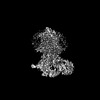
| Title | Structure of human Wnt7a bound to WLS and CALR | |||||||||||||||||||||||||||||||||||||||||||||
 Components Components |
| |||||||||||||||||||||||||||||||||||||||||||||
 Keywords Keywords | SIGNALING PROTEIN | |||||||||||||||||||||||||||||||||||||||||||||
| Function / homology |  Function and homology information Function and homology informationpostsynapse assembly / positive regulation of excitatory synapse assembly / positive regulation of protein localization to presynapse / skeletal muscle satellite cell activation / regulation of axon diameter / Wnt protein secretion / asymmetric protein localization involved in cell fate determination / response to biphenyl / Calnexin/calreticulin cycle / cytolytic granule ...postsynapse assembly / positive regulation of excitatory synapse assembly / positive regulation of protein localization to presynapse / skeletal muscle satellite cell activation / regulation of axon diameter / Wnt protein secretion / asymmetric protein localization involved in cell fate determination / response to biphenyl / Calnexin/calreticulin cycle / cytolytic granule / positive regulation of Wnt protein secretion / lens fiber cell development / cerebellar granule cell differentiation / WNT ligand biogenesis and trafficking / positive regulation of dendritic cell chemotaxis / nuclear receptor-mediated glucocorticoid signaling pathway / oviduct development / Assembly of Viral Components at the Budding Site / synaptic vesicle recycling / central nervous system vasculogenesis / ATF6 (ATF6-alpha) activates chaperone genes / cell proliferation in forebrain / excitatory synapse assembly / negative regulation of trophoblast cell migration / cortical granule / uterus morphogenesis / cellular response to electrical stimulus / response to peptide / embryonic axis specification / regulation of meiotic nuclear division / cementum mineralization / negative regulation of retinoic acid receptor signaling pathway / secondary palate development / sequestering of calcium ion / complement component C1q complex binding / presynapse assembly / somatic stem cell division / skeletal muscle satellite cell maintenance involved in skeletal muscle regeneration / endoplasmic reticulum quality control compartment / protein folding in endoplasmic reticulum / sarcoplasmic reticulum lumen / stem cell development / sex differentiation / hindbrain development / positive regulation of epithelial cell proliferation involved in wound healing / Wnt-protein binding / negative regulation of intracellular steroid hormone receptor signaling pathway / nuclear export signal receptor activity / cardiac muscle cell differentiation / establishment of blood-brain barrier / dendritic spine morphogenesis / frizzled binding / exocrine pancreas development / dorsal/ventral pattern formation / neurotransmitter secretion / embryonic forelimb morphogenesis / Class B/2 (Secretin family receptors) / regulation of postsynapse organization / embryonic hindlimb morphogenesis / positive regulation of synapse assembly / anterior/posterior axis specification / cortical actin cytoskeleton organization / wound healing, spreading of epidermal cells / Wnt signaling pathway, planar cell polarity pathway / response to glycoside / Scavenging by Class A Receptors / embryonic digit morphogenesis / nuclear androgen receptor binding / cartilage condensation / Scavenging by Class F Receptors / cellular response to lithium ion / negative regulation of neuron differentiation / response to testosterone / midbrain development / establishment of cell polarity / regulation of synaptic vesicle exocytosis / organelle membrane / positive regulation of protein metabolic process / smooth endoplasmic reticulum / hormone binding / mesoderm formation / molecular sequestering activity / somatic stem cell population maintenance / cell fate commitment / positive regulation of excitatory postsynaptic potential / chondrocyte differentiation / protein localization to nucleus / canonical Wnt signaling pathway / positive regulation of Wnt signaling pathway / regulation of presynapse assembly / cellular response to transforming growth factor beta stimulus / positive regulation of cell cycle / ERAD pathway / endomembrane system / endocytic vesicle lumen / endoplasmic reticulum-Golgi intermediate compartment membrane / protein folding chaperone / positive regulation of substrate adhesion-dependent cell spreading / extracellular matrix / peptide binding Similarity search - Function | |||||||||||||||||||||||||||||||||||||||||||||
| Biological species |  Homo sapiens (human) Homo sapiens (human) | |||||||||||||||||||||||||||||||||||||||||||||
| Method | ELECTRON MICROSCOPY / single particle reconstruction / cryo EM / Resolution: 3.1 Å | |||||||||||||||||||||||||||||||||||||||||||||
 Authors Authors | Qi, X. / Hu, Q. / Li, X. | |||||||||||||||||||||||||||||||||||||||||||||
| Funding support |  United States, 3items United States, 3items
| |||||||||||||||||||||||||||||||||||||||||||||
 Citation Citation | Journal: Cell / Year: 2023 Title: Molecular basis of Wnt biogenesis, secretion, and Wnt7-specific signaling. Authors: Xiaofeng Qi / Qinli Hu / Nadia Elghobashi-Meinhardt / Tao Long / Hongwen Chen / Xiaochun Li /  Abstract: Wnt proteins are enzymatically lipidated by Porcupine (PORCN) in the ER and bind to Wntless (WLS) for intracellular transport and secretion. Mechanisms governing the transfer of these low-solubility ...Wnt proteins are enzymatically lipidated by Porcupine (PORCN) in the ER and bind to Wntless (WLS) for intracellular transport and secretion. Mechanisms governing the transfer of these low-solubility Wnts from the ER to the extracellular space remain unclear. Through structural and functional analyses of Wnt7a, a crucial Wnt involved in central nervous system angiogenesis and blood-brain barrier maintenance, we have elucidated the principles of Wnt biogenesis and Wnt7-specific signaling. The Wnt7a-WLS complex binds to calreticulin (CALR), revealing that CALR functions as a chaperone to facilitate Wnt transfer from PORCN to WLS during Wnt biogenesis. Our structures, functional analyses, and molecular dynamics simulations demonstrate that a phospholipid in the core of Wnt-bound WLS regulates the association and dissociation between Wnt and WLS, suggesting a lipid-mediated Wnt secretion mechanism. Finally, the structure of Wnt7a bound to RECK, a cell-surface Wnt7 co-receptor, reveals how RECK engages the N-terminal domain of Wnt7a to activate Wnt7-specific signaling. | |||||||||||||||||||||||||||||||||||||||||||||
| History |
|
- Structure visualization
| Structure viewer | Molecule: MolmilJmol/JSmol |
|---|
- Downloads & links
-Download
| PDBx/mmCIF format | 8tzo.cif.gz | 237.1 KB | Display | PDBx/mmCIF format |
|---|---|---|---|---|
| PDB format | pdb8tzo.ent.gz | 181.7 KB | Display | PDB format |
| PDBx/mmJSON format | 8tzo.json.gz | Tree view | PDBx/mmJSON format | |
| Others |  Other downloads Other downloads |
-Validation report
| Summary document | 8tzo_validation.pdf.gz | 1.4 MB | Display | wwPDB validaton report |
|---|---|---|---|---|
| Full document | 8tzo_full_validation.pdf.gz | 1.4 MB | Display | |
| Data in XML | 8tzo_validation.xml.gz | 42.9 KB | Display | |
| Data in CIF | 8tzo_validation.cif.gz | 62 KB | Display | |
| Arichive directory | https://data.pdbj.org/pub/pdb/validation_reports/tz/8tzoftp://data.pdbj.org/pub/pdb/validation_reports/tz/8tzo | HTTPS FTP |
-Related structure data
| Related structure data |  41764MC 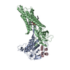 8tzpC 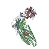 8tzrC  8tzsC M: map data used to model this data C: citing same article ( |
|---|---|
| Similar structure data |
-Links
 PDBj
PDBj


- Assembly
| Deposited unit | 
|
|---|---|
| 1 |
|
-Components
-Protein , 3 types, 3 molecules ABC
| #1: Protein | Mass: 39062.977 Da / Num. of mol.: 1 Source method: isolated from a genetically manipulated source Source: (gene. exp.) Homo sapiens (human) / Gene: WNT7A / Production host: Homo sapiens (human) / References: UniProt: O00755 |
|---|---|
| #2: Protein | Mass: 62317.973 Da / Num. of mol.: 1 Source method: isolated from a genetically manipulated source Source: (gene. exp.) Homo sapiens (human) / Gene: WLS / Production host: Homo sapiens (human) / References: UniProt: Q5T9L3 |
| #3: Protein | Mass: 48198.379 Da / Num. of mol.: 1 / Source method: isolated from a natural source / Source: (natural) Homo sapiens (human) / References: UniProt: P27797 |
-Sugars , 1 types, 1 molecules
| #4: Polysaccharide | alpha-D-glucopyranose-(1-3)-alpha-D-mannopyranose-(1-2)-alpha-D-mannopyranose-(1-2)-alpha-D- ...alpha-D-glucopyranose-(1-3)-alpha-D-mannopyranose-(1-2)-alpha-D-mannopyranose-(1-2)-alpha-D-mannopyranose-(1-3)-beta-D-mannopyranose-(1-4)-2-acetamido-2-deoxy-beta-D-glucopyranose-(1-4)-2-acetamido-2-deoxy-beta-D-glucopyranose Type: oligosaccharide / Mass: 1235.105 Da / Num. of mol.: 1 Source method: isolated from a genetically manipulated source |
|---|
-Non-polymers , 3 types, 3 molecules 


| #5: Chemical | ChemComp-PAM /  Mass: 254.408 Da / Num. of mol.: 1 / Source method: obtained synthetically / Formula: C16H30O2 / Feature type: SUBJECT OF INVESTIGATION Mass: 254.408 Da / Num. of mol.: 1 / Source method: obtained synthetically / Formula: C16H30O2 / Feature type: SUBJECT OF INVESTIGATION |
|---|---|
| #6: Chemical | ChemComp-POV / ( Mass: 760.076 Da / Num. of mol.: 1 / Source method: obtained synthetically / Formula: C42H82NO8P / Feature type: SUBJECT OF INVESTIGATION / Comment: phospholipid*YM Mass: 760.076 Da / Num. of mol.: 1 / Source method: obtained synthetically / Formula: C42H82NO8P / Feature type: SUBJECT OF INVESTIGATION / Comment: phospholipid*YM |
| #7: Chemical | ChemComp-CA /  Mass: 40.078 Da / Num. of mol.: 1 / Source method: obtained synthetically / Formula: Ca Mass: 40.078 Da / Num. of mol.: 1 / Source method: obtained synthetically / Formula: Ca |
-Details
| Has ligand of interest | Y |
|---|---|
| Has protein modification | Y |
-Experimental details
-Experiment
| Experiment | Method: ELECTRON MICROSCOPY |
|---|---|
| EM experiment | Aggregation state: PARTICLE / 3D reconstruction method: single particle reconstruction |
- Sample preparation
| Component | Name: Wnt7a-WLS-CALR Complex / Type: COMPLEX / Entity ID: #1-#3 / Source: MULTIPLE SOURCES |
|---|---|
| Source (natural) | Organism: Homo sapiens (human) |
| Source (recombinant) | Organism: Homo sapiens (human) |
| Buffer solution | pH: 7.5 |
| Specimen | Embedding applied: NO / Shadowing applied: NO / Staining applied: NO / Vitrification applied: YES |
| Vitrification | Cryogen name: ETHANE |
- Electron microscopy imaging
| Experimental equipment |  Model: Titan Krios / Image courtesy: FEI Company |
|---|---|
| Microscopy | Model: FEI TITAN KRIOS |
| Electron gun | Electron source:  FIELD EMISSION GUN / Accelerating voltage: 300 kV / Illumination mode: FLOOD BEAM FIELD EMISSION GUN / Accelerating voltage: 300 kV / Illumination mode: FLOOD BEAM |
| Electron lens | Mode: BRIGHT FIELD / Nominal defocus max: 2000 nm / Nominal defocus min: 1000 nm |
| Image recording | Electron dose: 60 e/Å2 / Film or detector model: GATAN K3 (6k x 4k) |
- Processing
| EM software | Name: PHENIX / Category: model refinement | ||||||||||||||||||||||||
|---|---|---|---|---|---|---|---|---|---|---|---|---|---|---|---|---|---|---|---|---|---|---|---|---|---|
| CTF correction | Type: NONE | ||||||||||||||||||||||||
| 3D reconstruction | Resolution: 3.1 Å / Resolution method: FSC 0.143 CUT-OFF / Num. of particles: 276377 / Symmetry type: POINT | ||||||||||||||||||||||||
| Refine LS restraints |
|
