- PDB-8t33: Crystal structure of K46 acetylated GABARAP in complex with the L... -
+
Open data
ID or keywords:
Loading...
-
Basic information
Entry
Database: PDB / ID: 8t33
Title
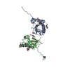
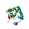
Crystal structure of K46 acetylated GABARAP in complex with the LIR of TP53INP2/DOR
Components
Gamma-aminobutyric acid receptor-associated protein
Tumor protein p53-inducible nuclear protein 2
Keywords
PROTEIN BINDING / Autophagy / GABARAP acetylation / DOR LIR / TP53INP2 LIR
Function / homology
Function and homology information
negative regulation of protein localization / positive regulation of protein K48-linked ubiquitination / regulation of Rac protein signal transduction / tissue homeostasis / GABA receptor binding / phosphatidylethanolamine binding / TBC/RABGAPs / cellular response to nitrogen starvation / microtubule associated complex / Macroautophagy ...negative regulation of protein localization / positive regulation of protein K48-linked ubiquitination / regulation of Rac protein signal transduction / tissue homeostasis / GABA receptor binding / phosphatidylethanolamine binding / TBC/RABGAPs / cellular response to nitrogen starvation / microtubule associated complex / Macroautophagy / regulation of neurotransmitter receptor localization to postsynaptic specialization membrane / smooth endoplasmic reticulum / autophagosome membrane / extrinsic apoptotic signaling pathway via death domain receptors / autophagosome maturation / axoneme / autophagosome assembly / beta-tubulin binding / mitophagy / protein targeting / autophagosome / ubiquitin binding / PML body / microtubule cytoskeleton organization / GABA-ergic synapse / osteoblast differentiation / intracellular protein localization / positive regulation of proteasomal ubiquitin-dependent protein catabolic process / protein transport / actin cytoskeleton / cytoplasmic vesicle / sperm midpiece / microtubule binding / ubiquitin-dependent protein catabolic process / microtubule / chemical synaptic transmission / lysosome / Golgi membrane / ubiquitin protein ligase binding / positive regulation of DNA-templated transcription / nucleus / plasma membrane / cytosol Similarity search - Function
Tumour protein p53-inducible nuclear protein / DOR family / Autophagy protein Atg8 ubiquitin-like / Autophagy protein Atg8 ubiquitin like / Ubiquitin-like domain superfamily Similarity search - Domain/homology
ACETATE ION / Gamma-aminobutyric acid receptor-associated protein / Tumor protein p53-inducible nuclear protein 2 Similarity search - Component
In the structure databanks used in Yorodumi, some data are registered as the other names, "COVID-19 virus" and "2019-nCoV". Here are the details of the virus and the list of structure data.
Jan 31, 2019. EMDB accession codes are about to change! (news from PDBe EMDB page)
EMDB accession codes are about to change! (news from PDBe EMDB page)
The allocation of 4 digits for EMDB accession codes will soon come to an end. Whilst these codes will remain in use, new EMDB accession codes will include an additional digit and will expand incrementally as the available range of codes is exhausted. The current 4-digit format prefixed with “EMD-” (i.e. EMD-XXXX) will advance to a 5-digit format (i.e. EMD-XXXXX), and so on. It is currently estimated that the 4-digit codes will be depleted around Spring 2019, at which point the 5-digit format will come into force.
The EM Navigator/Yorodumi systems omit the EMD- prefix.
Related info.:Q: What is EMD? / ID/Accession-code notation in Yorodumi/EM Navigator
Yorodumi is a browser for structure data from EMDB, PDB, SASBDB, etc.
This page is also the successor to EM Navigator detail page, and also detail information page/front-end page for Omokage search.
The word "yorodu" (or yorozu) is an old Japanese word meaning "ten thousand". "mi" (miru) is to see.
Related info.:EMDB / PDB / SASBDB / Comparison of 3 databanks / Yorodumi Search / Aug 31, 2016. New EM Navigator & Yorodumi / Yorodumi Papers / Jmol/JSmol / Function and homology information / Changes in new EM Navigator and Yorodumi
 Movie
Movie Controller

 Yorodumi
 Open data
 Basic information
 Components
 Keywords
 Function and homology information
 Homo sapiens (human)
 X-RAY DIFFRACTION /
  Authors
 Canada, 1items
  Citation
 Structure visualization
 Downloads & links
 Other downloads
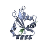


 PDBj
PDBj


 Assembly


 Mass: 65.409 Da / Num. of mol.: 9 / Source method: obtained synthetically / Formula: Zn / Feature type: SUBJECT OF INVESTIGATION
 Mass: 59.044 Da / Num. of mol.: 4 / Source method: obtained synthetically / Formula: C2H3O2 / Feature type: SUBJECT OF INVESTIGATION
 Mass: 18.015 Da / Num. of mol.: 200 / Source method: isolated from a natural source / Formula: H2O
 Sample preparation
 Processing