[English] 日本語
Yorodumi
- PDB-8t2l: Crystal structure of LC3A in complex with the LIR of NSs4 -

+
Open data


ID or keywords:

Loading...

-
Basic information

Entry
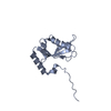
Database: PDB / ID: 8t2l
TitleCrystal structure of LC3A in complex with the LIR of NSs4
ComponentsNon-structural protein S,Microtubule-associated proteins 1A/1B light chain 3A chimera
KeywordsPROTEIN BINDING / Rift Valley fever virus / autophagy / LC3 / GABARAP / LIR
Function / homology
Function and homology information


symbiont-mediated suppression of host transcription initiation from RNA polymerase II promoter / symbiont-mediated suppression of host transcription / cellular response to oxygen-glucose deprivation / suppression by virus of host type I interferon production / symbiont-mediated suppression of host PKR/eIFalpha signaling / autophagy of mitochondrion / cellular response to nitrogen starvation / SMAD protein signal transduction / negative stranded viral RNA replication / phosphatidylethanolamine binding ...symbiont-mediated suppression of host transcription initiation from RNA polymerase II promoter / symbiont-mediated suppression of host transcription / cellular response to oxygen-glucose deprivation / suppression by virus of host type I interferon production / symbiont-mediated suppression of host PKR/eIFalpha signaling / autophagy of mitochondrion / cellular response to nitrogen starvation / SMAD protein signal transduction / negative stranded viral RNA replication / phosphatidylethanolamine binding / protein serine/threonine kinase inhibitor activity / response to iron(II) ion / autolysosome / Macroautophagy / Receptor Mediated Mitophagy / p38MAPK cascade / organelle membrane / autophagosome membrane / autophagosome maturation / autophagosome assembly / autophagosome / JNK cascade / cellular response to copper ion / PINK1-PRKN Mediated Mitophagy / cellular response to amino acid starvation / cellular response to starvation / macroautophagy / response to lead ion / phospholipid binding / cellular response to hydrogen peroxide / late endosome / symbiont-mediated suppression of host gene expression / microtubule binding / microtubule / host cell cytoplasm / symbiont-mediated suppression of host type I interferon-mediated signaling pathway / intracellular membrane-bounded organelle / glutamatergic synapse / ubiquitin protein ligase binding / host cell nucleus / cytosol
Similarity search - Function
Phlebovirus, non structural protein / Rift valley fever virus non structural protein-like / Rift valley fever virus non structural protein (NSs) like / Autophagy protein Atg8 ubiquitin-like / Autophagy protein Atg8 ubiquitin like / Ubiquitin-like domain superfamily
Similarity search - Domain/homology
Non-structural protein S / Microtubule-associated proteins 1A/1B light chain 3A
Similarity search - Component
Biological speciesHomo sapiens (human)
MethodX-RAY DIFFRACTION / SYNCHROTRON / MOLECULAR REPLACEMENT / Resolution: 2.24 Å
AuthorsAli, M.G.H. / Wahba, H.M. / Cyr, N. / Omichinski, J.G.
Funding support United States, 1items
OrganizationGrant numberCountry
Defense Threat Reduction Agency (DTRA) United States
CitationJournal: Plos Pathog. / Year: 2024
Title: An LIR motif in the Rift Valley fever virus NSs protein is critical for the interaction with LC3 family members and inhibition of autophagy.
Authors: Petraccione, K. / Ali, M.G.H. / Cyr, N. / Wahba, H.M. / Stocker, T. / Akhrymuk, M. / Akhrymuk, I. / Panny, L. / Bracci, N. / Cafaro, R. / Sastre, D. / Silberfarb, A. / O'Maille, P. / ...Authors: Petraccione, K. / Ali, M.G.H. / Cyr, N. / Wahba, H.M. / Stocker, T. / Akhrymuk, M. / Akhrymuk, I. / Panny, L. / Bracci, N. / Cafaro, R. / Sastre, D. / Silberfarb, A. / O'Maille, P. / Omichinski, J. / Kehn-Hall, K.
History
DepositionJun 6, 2023Deposition site: RCSB / Processing site: RCSB
Revision 1.0Apr 3, 2024Provider: repository / Type: Initial release

-
Structure visualization

Structure viewerMolecule:
MolmilJmol/JSmol

Downloads & links

-
Assembly

Deposited unit
A: Non-structural protein S,Microtubule-associated proteins 1A/1B light chain 3A chimera
B: Non-structural protein S,Microtubule-associated proteins 1A/1B light chain 3A chimera


Theoretical massNumber of molelcules
Total (without water)30,8592
Polymers30,8592
Non-polymers00
Water1,00956
1
A: Non-structural protein S,Microtubule-associated proteins 1A/1B light chain 3A chimera


Theoretical massNumber of molelcules
Total (without water)15,4301
Polymers15,4301
Non-polymers00
Water181
TypeNameSymmetry operationNumber
identity operation1_555x,y,z1
2
B: Non-structural protein S,Microtubule-associated proteins 1A/1B light chain 3A chimera


Theoretical massNumber of molelcules
Total (without water)15,4301
Polymers15,4301
Non-polymers00
Water181
TypeNameSymmetry operationNumber
identity operation1_555x,y,z1
Unit cell
Length a, b, c (Å)36.995, 50.267, 137.874
Angle α, β, γ (deg.)90.00, 90.00, 90.00
Int Tables number19
Space group name H-MP212121

-
Components

#1: Protein Non-structural protein S,Microtubule-associated proteins 1A/1B light chain 3A chimera / NSs / Autophagy-related protein LC3 A / Autophagy-related ubiquitin-like modifier LC3 A / MAP1 ...NSs / Autophagy-related protein LC3 A / Autophagy-related ubiquitin-like modifier LC3 A / MAP1 light chain 3-like protein 1 / MAP1A/MAP1B light chain 3 A / MAP1A/MAP1B LC3 A / Microtubule-associated protein 1 light chain 3 alpha


Mass: 15429.536 Da / Num. of mol.: 2
Fragment: GS is the remain of the removed expression tag. The N terminus of Nss has been fused with Q9H492 to facilitate the crystalization.
Source method: isolated from a genetically manipulated source
Source: (gene. exp.) Homo sapiens (human) / Gene: NSS, MAP1LC3A / Production host: Escherichia coli BL21(DE3) (bacteria) / References: UniProt: P21698, UniProt: Q9H492
#2: Water ChemComp-HOH / water


Mass: 18.015 Da / Num. of mol.: 56 / Source method: isolated from a natural source / Formula: H2O

-
Experimental details

-
Experiment

ExperimentMethod: X-RAY DIFFRACTION / Number of used crystals: 1

-
Sample preparation

CrystalDensity Matthews: 2.08 Å3/Da / Density % sol: 40.78 %
Crystal growTemperature: 293 K / Method: vapor diffusion, hanging drop
Details: 20% PEG3350, 0.2M Na-citrate, 5% Isopropanol, 0.1M MES buffer PH 6.5

-
Data collection

DiffractionMean temperature: 100 K / Serial crystal experiment: N
Diffraction sourceSource: SYNCHROTRON / Site: CHESS / Beamline: F1 / Wavelength: 0.9768 Å
DetectorType: DECTRIS PILATUS3 6M / Detector: PIXEL / Date: Mar 1, 2017
RadiationProtocol: SINGLE WAVELENGTH / Monochromatic (M) / Laue (L): M / Scattering type: x-ray
Radiation wavelengthWavelength: 0.9768 Å / Relative weight: 1
ReflectionResolution: 2.24→32.6 Å / Num. obs: 12899 / % possible obs: 93.28 % / Redundancy: 6.2 % / CC1/2: 0.997 / Net I/σ(I): 10.9
Reflection shellResolution: 2.24→2.32 Å / Num. unique obs: 1072 / CC1/2: 0.747

-
Processing

Software
NameVersionClassification
PHENIX1.14_3260refinement
HKL-2000data reduction
HKL-2000data scaling
PHASERphasing
RefinementMethod to determine structure: MOLECULAR REPLACEMENT / Resolution: 2.24→32.6 Å / SU ML: 0.32 / Cross valid method: FREE R-VALUE / σ(F): 0.33 / Phase error: 29.2 / Stereochemistry target values: ML
RfactorNum. reflection% reflection
Rfree0.2762 1217 10.03 %
Rwork0.2308 --
obs0.2354 12135 93.32 %
Solvent computationShrinkage radii: 0.9 Å / VDW probe radii: 1.11 Å / Solvent model: FLAT BULK SOLVENT MODEL
Refinement stepCycle: LAST / Resolution: 2.24→32.6 Å
ProteinNucleic acidLigandSolventTotal
Num. atoms2086 0 0 56 2142
Refine LS restraints
Refine-IDTypeDev idealNumber
X-RAY DIFFRACTIONf_bond_d0.0022139
X-RAY DIFFRACTIONf_angle_d0.5272885
X-RAY DIFFRACTIONf_dihedral_angle_d11.638843
X-RAY DIFFRACTIONf_chiral_restr0.023313
X-RAY DIFFRACTIONf_plane_restr0.003380
LS refinement shell
Resolution (Å)Rfactor RfreeNum. reflection RfreeRfactor RworkNum. reflection RworkRefine-ID% reflection obs (%)
2.24-2.32950.36781180.30271075X-RAY DIFFRACTION85
2.3295-2.43550.35171180.28711102X-RAY DIFFRACTION87
2.4355-2.56390.32791260.29091142X-RAY DIFFRACTION89
2.5639-2.72440.30691330.26821177X-RAY DIFFRACTION93
2.7244-2.93470.27941370.25451211X-RAY DIFFRACTION94
2.9347-3.22980.3161390.25341245X-RAY DIFFRACTION97
3.2298-3.69670.30861440.23371287X-RAY DIFFRACTION97
3.6967-4.65550.2311470.1981305X-RAY DIFFRACTION99
4.6555-32.60.23941550.20071374X-RAY DIFFRACTION98
Refinement TLS params.

Method: refined / Refine-ID: X-RAY DIFFRACTION

IDL112)L122)L132)L222)L232)L332)S11 (Å °)S12 (Å °)S13 (Å °)S21 (Å °)S22 (Å °)S23 (Å °)S31 (Å °)S32 (Å °)S33 (Å °)T112)T122)T132)T222)T232)T332)Origin x (Å)Origin y (Å)Origin z (Å)
13.70870.3718-0.06271.13410.3893.2617-0.4003-0.01280.1752-0.52010.2102-0.286-0.6461-0.15140.08780.53720.062-0.07260.3354-0.00730.2543-11.1795-25.38399.0249
22.74510.20050.94672.6706-0.05211.5416-0.05670.05640.0192-0.01510.073-0.1026-0.14720.0436-0.02920.20450.0180.07290.2648-0.01660.2848-6.6295-8.933233.6577
Refinement TLS group
IDRefine-IDRefine TLS-IDSelection details
1X-RAY DIFFRACTION1chain B
2X-RAY DIFFRACTION2chain A

+
About Yorodumi

-
News

-
Feb 9, 2022. New format data for meta-information of EMDB entries

New format data for meta-information of EMDB entries

  • Version 3 of the EMDB header file is now the official format.
  • The previous official version 1.9 will be removed from the archive.

Related info.:EMDB header

External links:wwPDB to switch to version 3 of the EMDB data model

-
Aug 12, 2020. Covid-19 info

Covid-19 info

URL: https://pdbjlvh1.pdbj.org/emnavi/covid19.php

New page: Covid-19 featured information page in EM Navigator.

Related info.:Covid-19 info / Mar 5, 2020. Novel coronavirus structure data

+
Mar 5, 2020. Novel coronavirus structure data

Novel coronavirus structure data

Related info.:Yorodumi Speices / Aug 12, 2020. Covid-19 info

External links:COVID-19 featured content - PDBj / Molecule of the Month (242):Coronavirus Proteases

+
Jan 31, 2019. EMDB accession codes are about to change! (news from PDBe EMDB page)

EMDB accession codes are about to change! (news from PDBe EMDB page)

  • The allocation of 4 digits for EMDB accession codes will soon come to an end. Whilst these codes will remain in use, new EMDB accession codes will include an additional digit and will expand incrementally as the available range of codes is exhausted. The current 4-digit format prefixed with “EMD-” (i.e. EMD-XXXX) will advance to a 5-digit format (i.e. EMD-XXXXX), and so on. It is currently estimated that the 4-digit codes will be depleted around Spring 2019, at which point the 5-digit format will come into force.
  • The EM Navigator/Yorodumi systems omit the EMD- prefix.

Related info.:Q: What is EMD? / ID/Accession-code notation in Yorodumi/EM Navigator

External links:EMDB Accession Codes are Changing Soon! / Contact to PDBj

+
Jul 12, 2017. Major update of PDB

Major update of PDB

  • wwPDB released updated PDB data conforming to the new PDBx/mmCIF dictionary.
  • This is a major update changing the version number from 4 to 5, and with Remediation, in which all the entries are updated.
  • In this update, many items about electron microscopy experimental information are reorganized (e.g. em_software).
  • Now, EM Navigator and Yorodumi are based on the updated data.

External links:wwPDB Remediation / Enriched Model Files Conforming to OneDep Data Standards Now Available in the PDB FTP Archive

-
Yorodumi

Thousand views of thousand structures

  • Yorodumi is a browser for structure data from EMDB, PDB, SASBDB, etc.
  • This page is also the successor to EM Navigator detail page, and also detail information page/front-end page for Omokage search.
  • The word "yorodu" (or yorozu) is an old Japanese word meaning "ten thousand". "mi" (miru) is to see.

Related info.:EMDB / PDB / SASBDB / Comparison of 3 databanks / Yorodumi Search / Aug 31, 2016. New EM Navigator & Yorodumi / Yorodumi Papers / Jmol/JSmol / Function and homology information / Changes in new EM Navigator and Yorodumi

Read more