 Movie
Movie Controller

[English] 日本語
 Yorodumi
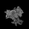
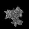
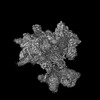
- PDB-8rg0: Structure of human eIF3 core from closed 48S translation initiati... -
+ Open data

- Basic information
| Entry | Database: PDB / ID: 8rg0 | |||||||||||||||||||||||||||||||||||||||||||||||||||||||||||||||||||||||||||||||||||||||||||||||||||||||||
|---|---|---|---|---|---|---|---|---|---|---|---|---|---|---|---|---|---|---|---|---|---|---|---|---|---|---|---|---|---|---|---|---|---|---|---|---|---|---|---|---|---|---|---|---|---|---|---|---|---|---|---|---|---|---|---|---|---|---|---|---|---|---|---|---|---|---|---|---|---|---|---|---|---|---|---|---|---|---|---|---|---|---|---|---|---|---|---|---|---|---|---|---|---|---|---|---|---|---|---|---|---|---|---|---|---|---|
| Title | Structure of human eIF3 core from closed 48S translation initiation complex | |||||||||||||||||||||||||||||||||||||||||||||||||||||||||||||||||||||||||||||||||||||||||||||||||||||||||
 Components Components |
| |||||||||||||||||||||||||||||||||||||||||||||||||||||||||||||||||||||||||||||||||||||||||||||||||||||||||
 Keywords Keywords | RIBOSOME / TRANSLATION / initiation / 48S / eIF / human / eukaryotic / factor / codon / scanning / open / reading | |||||||||||||||||||||||||||||||||||||||||||||||||||||||||||||||||||||||||||||||||||||||||||||||||||||||||
| Function / homology |  Function and homology information Function and homology informationviral translational termination-reinitiation / eukaryotic translation initiation factor 3 complex, eIF3e / cap-dependent translational initiation / eukaryotic translation initiation factor 3 complex, eIF3m / IRES-dependent viral translational initiation / translation reinitiation / formation of cytoplasmic translation initiation complex / multi-eIF complex / eukaryotic translation initiation factor 3 complex / eukaryotic 43S preinitiation complex ...viral translational termination-reinitiation / eukaryotic translation initiation factor 3 complex, eIF3e / cap-dependent translational initiation / eukaryotic translation initiation factor 3 complex, eIF3m / IRES-dependent viral translational initiation / translation reinitiation / formation of cytoplasmic translation initiation complex / multi-eIF complex / eukaryotic translation initiation factor 3 complex / eukaryotic 43S preinitiation complex / mRNA cap binding / eukaryotic 48S preinitiation complex / nuclear-transcribed mRNA catabolic process, nonsense-mediated decay / regulation of translational initiation / negative regulation of RNA splicing / ZNF598 and the Ribosome-associated Quality Trigger (RQT) complex dissociate a ribosome stalled on a no-go mRNA / metal-dependent deubiquitinase activity / PELO:HBS1L and ABCE1 dissociate a ribosome on a non-stop mRNA / cytoplasmic translational initiation / rRNA modification in the nucleus and cytosol / erythrocyte homeostasis / cytoplasmic side of rough endoplasmic reticulum membrane / Formation of the ternary complex, and subsequently, the 43S complex / laminin receptor activity / Ribosomal scanning and start codon recognition / Translation initiation complex formation / SARS-CoV-1 modulates host translation machinery / Peptide chain elongation / Selenocysteine synthesis / Formation of a pool of free 40S subunits / Eukaryotic Translation Termination / SRP-dependent cotranslational protein targeting to membrane / Response of EIF2AK4 (GCN2) to amino acid deficiency / Viral mRNA Translation / Nonsense Mediated Decay (NMD) independent of the Exon Junction Complex (EJC) / GTP hydrolysis and joining of the 60S ribosomal subunit / L13a-mediated translational silencing of Ceruloplasmin expression / Major pathway of rRNA processing in the nucleolus and cytosol / Nonsense Mediated Decay (NMD) enhanced by the Exon Junction Complex (EJC) / Amplification of signal from unattached kinetochores via a MAD2 inhibitory signal / laminin binding / translation initiation factor activity / negative regulation of proteasomal ubiquitin-dependent protein catabolic process / negative regulation of translational initiation / Mitotic Prometaphase / translation initiation factor binding / EML4 and NUDC in mitotic spindle formation / Resolution of Sister Chromatid Cohesion / antiviral innate immune response / erythrocyte differentiation / cytosolic ribosome / positive regulation of translation / maturation of SSU-rRNA / translational initiation / small-subunit processome / negative regulation of ERK1 and ERK2 cascade / RHO GTPases Activate Formins / receptor tyrosine kinase binding / PML body / fibrillar center / GABA-ergic synapse / mRNA 5'-UTR binding / Regulation of expression of SLITs and ROBOs / metallopeptidase activity / rRNA processing / Separation of Sister Chromatids / ribosomal small subunit assembly / presynapse / virus receptor activity / ribosome binding / ribosome biogenesis / ribosomal small subunit biogenesis / small ribosomal subunit / small ribosomal subunit rRNA binding / cytosolic small ribosomal subunit / microtubule / SARS-CoV-2 modulates host translation machinery / cytoplasmic translation / cell differentiation / cysteine-type deubiquitinase activity / ubiquitinyl hydrolase 1 / postsynaptic density / structural constituent of ribosome / cadherin binding / ribosome / translation / ribonucleoprotein complex / focal adhesion / mRNA binding / synapse / negative regulation of apoptotic process / nucleolus / chromatin / glutamatergic synapse / structural molecule activity / negative regulation of transcription by RNA polymerase II / endoplasmic reticulum / proteolysis / DNA binding / RNA binding Similarity search - Function | |||||||||||||||||||||||||||||||||||||||||||||||||||||||||||||||||||||||||||||||||||||||||||||||||||||||||
| Biological species |  Homo sapiens (human) Homo sapiens (human) | |||||||||||||||||||||||||||||||||||||||||||||||||||||||||||||||||||||||||||||||||||||||||||||||||||||||||
| Method | ELECTRON MICROSCOPY / single particle reconstruction / cryo EM / Resolution: 3.4 Å | |||||||||||||||||||||||||||||||||||||||||||||||||||||||||||||||||||||||||||||||||||||||||||||||||||||||||
 Authors Authors | Petrychenko, V. / Yi, S.-H. / Liedtke, D. / Peng, B.Z. / Rodnina, M.V. / Fischer, N. | |||||||||||||||||||||||||||||||||||||||||||||||||||||||||||||||||||||||||||||||||||||||||||||||||||||||||
| Funding support |  Germany, 2items Germany, 2items
| |||||||||||||||||||||||||||||||||||||||||||||||||||||||||||||||||||||||||||||||||||||||||||||||||||||||||
 Citation Citation | Journal: Nat Struct Mol Biol / Year: 2025 Title: Structural basis for translational control by the human 48S initiation complex. Authors: Valentyn Petrychenko / Sung-Hui Yi / David Liedtke / Bee-Zen Peng / Marina V Rodnina / Niels Fischer / Abstract: The selection of an open reading frame (ORF) for translation of eukaryotic mRNA relies on remodeling of the scanning 48S initiation complex into an elongation-ready 80S ribosome. Using cryo-electron ...The selection of an open reading frame (ORF) for translation of eukaryotic mRNA relies on remodeling of the scanning 48S initiation complex into an elongation-ready 80S ribosome. Using cryo-electron microscopy, we visualize the key commitment steps orchestrating 48S remodeling in humans. The mRNA Kozak sequence facilitates mRNA scanning in the 48S open state and stabilizes the 48S closed state by organizing the contacts of eukaryotic initiation factors (eIFs) and ribosomal proteins and by reconfiguring mRNA structure. GTPase-triggered large-scale fluctuations of 48S-bound eIF2 facilitate eIF5B recruitment, transfer of initiator tRNA from eIF2 to eIF5B and the release of eIF5 and eIF2. The 48S-bound multisubunit eIF3 complex controls ribosomal subunit joining by coupling eIF exchange to gradual displacement of the eIF3c N-terminal domain from the intersubunit interface. These findings reveal the structural mechanism of ORF selection in human cells and explain how eIF3 could function in the context of the 80S ribosome. | |||||||||||||||||||||||||||||||||||||||||||||||||||||||||||||||||||||||||||||||||||||||||||||||||||||||||
| History |
|
- Structure visualization
| Structure viewer | Molecule: MolmilJmol/JSmol |
|---|
- Downloads & links
-Download
| PDBx/mmCIF format | 8rg0.cif.gz | 833 KB | Display | PDBx/mmCIF format |
|---|---|---|---|---|
| PDB format | pdb8rg0.ent.gz | 593.6 KB | Display | PDB format |
| PDBx/mmJSON format | 8rg0.json.gz | Tree view | PDBx/mmJSON format | |
| Others |  Other downloads Other downloads |
-Validation report
| Arichive directory | https://data.pdbj.org/pub/pdb/validation_reports/rg/8rg0ftp://data.pdbj.org/pub/pdb/validation_reports/rg/8rg0 | HTTPS FTP |
|---|
-Related structure data
| Related structure data | 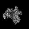 19128MC  8pj1C  8pj2C  8pj3C  8pj4C  8pj5C  8pj6C C: citing same article ( M: map data used to model this data |
|---|---|
| Similar structure data |


-Links
 PDBj
PDBj


- Assembly
| Deposited unit | 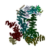
|
|---|---|
| 1 |
|
-Components
-Eukaryotic translation initiation factor 3 subunit ... , 9 types, 9 molecules 34568uvxy
| #1: Protein | Mass: 25083.619 Da / Num. of mol.: 1 / Source method: isolated from a natural source / Source: (natural) Homo sapiens (human) / References: UniProt: Q9UBQ5 |
|---|---|
| #2: Protein | Mass: 37593.645 Da / Num. of mol.: 1 / Source method: isolated from a natural source / Source: (natural) Homo sapiens (human) / References: UniProt: O00303, ubiquitinyl hydrolase 1 |
| #3: Protein | Mass: 66803.734 Da / Num. of mol.: 1 / Source method: isolated from a natural source / Source: (natural) Homo sapiens (human) / References: UniProt: Q9Y262 |
| #4: Protein | Mass: 42555.832 Da / Num. of mol.: 1 / Source method: isolated from a natural source / Source: (natural) Homo sapiens (human) / References: UniProt: Q7L2H7 |
| #6: Protein | Mass: 39979.277 Da / Num. of mol.: 1 / Source method: isolated from a natural source / Source: (natural) Homo sapiens (human) / References: UniProt: O15372 |
| #16: Protein | Mass: 166903.781 Da / Num. of mol.: 1 / Source method: isolated from a natural source / Source: (natural) Homo sapiens (human) / References: UniProt: Q14152 |
| #17: Protein | Mass: 52281.633 Da / Num. of mol.: 1 / Source method: isolated from a natural source / Source: (natural) Homo sapiens (human) / References: UniProt: P60228 |
| #18: Protein | Mass: 64060.758 Da / Num. of mol.: 1 / Source method: isolated from a natural source / Source: (natural) Homo sapiens (human) / References: UniProt: O15371 |
| #19: Protein | Mass: 105503.945 Da / Num. of mol.: 1 / Source method: isolated from a natural source / Source: (natural) Homo sapiens (human) / References: UniProt: Q99613 |
-RNA chain , 2 types, 2 molecules 7A
| #5: RNA chain | Mass: 82412.445 Da / Num. of mol.: 1 Source method: isolated from a genetically manipulated source Source: (gene. exp.) Homo sapiens (human) / Production host:  |
|---|---|
| #7: RNA chain | Mass: 603245.500 Da / Num. of mol.: 1 / Source method: isolated from a natural source Details: Only small part of the 18S rRNA is provided with sequence truncated to visualized only parts. Source: (natural) Homo sapiens (human) |
-40S ribosomal protein ... , 8 types, 8 molecules HIMNOPQn
| #8: Protein | Mass: 9480.186 Da / Num. of mol.: 1 / Source method: isolated from a natural source / Source: (natural) Homo sapiens (human) / References: UniProt: P42677 |
|---|---|
| #9: Protein | Mass: 17259.389 Da / Num. of mol.: 1 / Source method: isolated from a natural source / Source: (natural) Homo sapiens (human) / References: UniProt: P62277 |
| #10: Protein | Mass: 15578.156 Da / Num. of mol.: 1 / Source method: isolated from a natural source / Source: (natural) Homo sapiens (human) / References: UniProt: P08708 |
| #11: Protein | Mass: 32883.938 Da / Num. of mol.: 1 / Source method: isolated from a natural source / Source: (natural) Homo sapiens (human) / References: UniProt: P08865 |
| #12: Protein | Mass: 30002.061 Da / Num. of mol.: 1 / Source method: isolated from a natural source / Source: (natural) Homo sapiens (human) / References: UniProt: P61247 |
| #13: Protein | Mass: 16302.772 Da / Num. of mol.: 1 / Source method: isolated from a natural source / Source: (natural) Homo sapiens (human) / References: UniProt: P62263 |
| #14: Protein | Mass: 13047.532 Da / Num. of mol.: 1 / Source method: isolated from a natural source / Source: (natural) Homo sapiens (human) / References: UniProt: P62854 |
| #15: Protein | Mass: 7855.052 Da / Num. of mol.: 1 / Source method: isolated from a natural source / Source: (natural) Homo sapiens (human) / References: UniProt: P62857 |
-Non-polymers , 2 types, 2 molecules 

| #20: Chemical | ChemComp-MG /  Mass: 24.305 Da / Num. of mol.: 1 / Source method: obtained synthetically / Formula: Mg Mass: 24.305 Da / Num. of mol.: 1 / Source method: obtained synthetically / Formula: Mg |
|---|---|
| #21: Chemical | ChemComp-ZN /  Mass: 65.409 Da / Num. of mol.: 1 / Source method: obtained synthetically / Formula: Zn Mass: 65.409 Da / Num. of mol.: 1 / Source method: obtained synthetically / Formula: Zn |
-Details
| Has ligand of interest | N |
|---|---|
| Has protein modification | Y |
-Experimental details
-Experiment
| Experiment | Method: ELECTRON MICROSCOPY |
|---|---|
| EM experiment | Aggregation state: PARTICLE / 3D reconstruction method: single particle reconstruction |
- Sample preparation
| Component |
| ||||||||||||||||||||||||||||||||||||||||
|---|---|---|---|---|---|---|---|---|---|---|---|---|---|---|---|---|---|---|---|---|---|---|---|---|---|---|---|---|---|---|---|---|---|---|---|---|---|---|---|---|---|
| Source (natural) |
| ||||||||||||||||||||||||||||||||||||||||
| Source (recombinant) | Organism: | ||||||||||||||||||||||||||||||||||||||||
| Buffer solution | pH: 7.5 | ||||||||||||||||||||||||||||||||||||||||
| Buffer component |
| ||||||||||||||||||||||||||||||||||||||||
| Specimen | Embedding applied: NO / Shadowing applied: NO / Staining applied: NO / Vitrification applied: YES | ||||||||||||||||||||||||||||||||||||||||
| Vitrification | Instrument: HOMEMADE PLUNGER / Cryogen name: ETHANE / Humidity: 100 % / Chamber temperature: 277 K / Details: Manual blotting & plunge-freezing |
- Electron microscopy imaging
| Experimental equipment |  Model: Titan Krios / Image courtesy: FEI Company |
|---|---|
| Microscopy | Model: FEI TITAN KRIOS |
| Electron gun | Electron source:  FIELD EMISSION GUN / Accelerating voltage: 300 kV / Illumination mode: SPOT SCAN FIELD EMISSION GUN / Accelerating voltage: 300 kV / Illumination mode: SPOT SCAN |
| Electron lens | Mode: BRIGHT FIELD / Nominal magnification: 59000 X / Nominal defocus max: 2500 nm / Nominal defocus min: 200 nm / Cs: 0.01 mm |
| Specimen holder | Cryogen: NITROGEN / Specimen holder model: FEI TITAN KRIOS AUTOGRID HOLDER |
| Image recording | Average exposure time: 1.5 sec. / Electron dose: 45 e/Å2 / Detector mode: INTEGRATING / Film or detector model: FEI FALCON III (4k x 4k) |
| EM imaging optics | Spherical aberration corrector: Electron-optical aberrations were corrected using a CETCOR Cs-corrector (CEOS, Heidelberg) aligned with the CETCORPLUS 4.6.9 software package (CEOS, Heidelberg). |
- Processing
| EM software |
| ||||||||||||
|---|---|---|---|---|---|---|---|---|---|---|---|---|---|
| CTF correction | Type: PHASE FLIPPING AND AMPLITUDE CORRECTION | ||||||||||||
| Symmetry | Point symmetry: C1 (asymmetric) | ||||||||||||
| 3D reconstruction | Resolution: 3.4 Å / Resolution method: FSC 0.143 CUT-OFF / Num. of particles: 356632 / Symmetry type: POINT | ||||||||||||
| Atomic model building | Space: REAL |
