 Movie
Movie Controller


+ Open data

- Basic information
| Entry | Database: PDB / ID: 8qpg | ||||||
|---|---|---|---|---|---|---|---|
| Title | Turret of Haloferax tailed virus 1 | ||||||
 Components Components |
| ||||||
 Keywords Keywords | VIRUS / Archeal virus / turret / turret capsid interface / Mg ions | ||||||
| Function / homology | NodB homology domain / Polysaccharide deacetylase / hydrolase activity, acting on carbon-nitrogen (but not peptide) bonds / Glycoside hydrolase/deacetylase, beta/alpha-barrel / carbohydrate metabolic process / Prokaryotic polysaccharide deacetylase Function and homology information Function and homology information | ||||||
| Biological species |  Haloferax tailed virus 1 Haloferax tailed virus 1 | ||||||
| Method | ELECTRON MICROSCOPY / single particle reconstruction / cryo EM / Resolution: 2.36 Å | ||||||
 Authors Authors | Zhang, D. / Daum, B. / Isupov, M.N. / McLaren, M. | ||||||
| Funding support | European Union, 1items
| ||||||
 Citation Citation | Journal: Sci Adv / Year: 2025 Title: Cryo-EM resolves the structure of the archaeal dsDNA virus HFTV1 from head to tail. Authors: Daniel X Zhang / Michail N Isupov / Rebecca M Davies / Sabine Schwarzer / Mathew McLaren / William S Stuart / Vicki A M Gold / Hanna M Oksanen / Tessa E F Quax / Bertram Daum /    Abstract: While archaeal viruses show a stunning diversity of morphologies, many bear a notable resemblance to tailed bacterial phages. This raises fundamental questions: Do all tailed viruses share a common ...While archaeal viruses show a stunning diversity of morphologies, many bear a notable resemblance to tailed bacterial phages. This raises fundamental questions: Do all tailed viruses share a common origin and do they infect their hosts in similar ways? Answering these questions requires high-resolution structural insights, yet no complete atomic models of archaeal viruses have been available. Here, we present the near-atomic resolution structure of Haloferax tailed virus 1 (HFTV1), an archaeal virus thriving in extreme salinity. Using cryo-electron microscopy, we resolve the architecture and assembly of all structural proteins and capture conformational transitions associated with DNA ejection. Our data reveal genome spooling within the capsid and identify putative receptor-binding and catalytic sites for host recognition and infection. These findings uncover key mechanisms of archaeal virus assembly, principles of virus-host interactions, and evolutionary links connecting archaeal, bacterial, and eukaryotic viruses. | ||||||
| History |
|
- Structure visualization
| Structure viewer | Molecule: MolmilJmol/JSmol |
|---|
- Downloads & links
-Download
| PDBx/mmCIF format | 8qpg.cif.gz | 360 KB | Display | PDBx/mmCIF format |
|---|---|---|---|---|
| PDB format | pdb8qpg.ent.gz | Display | PDB format | |
| PDBx/mmJSON format | 8qpg.json.gz | Tree view | PDBx/mmJSON format | |
| Others |  Other downloads Other downloads |
-Validation report
| Summary document | 8qpg_validation.pdf.gz | 1.1 MB | Display | wwPDB validaton report |
|---|---|---|---|---|
| Full document | 8qpg_full_validation.pdf.gz | 1.2 MB | Display | |
| Data in XML | 8qpg_validation.xml.gz | 58 KB | Display | |
| Data in CIF | 8qpg_validation.cif.gz | 90.2 KB | Display | |
| Arichive directory | https://data.pdbj.org/pub/pdb/validation_reports/qp/8qpgftp://data.pdbj.org/pub/pdb/validation_reports/qp/8qpg | HTTPS FTP |
-Related structure data
| Related structure data | 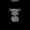 18550MC  8qpqC 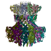 8qqnC  8qsiC  8qsyC  9fkbC  9gs0C  9h4pC  9h5bC  9h7vC M: map data used to model this data C: citing same article ( |
|---|---|
| Similar structure data |


-Links
 PDBj
PDBj
- Assembly
| Deposited unit | 
|
|---|---|
| 1 |
|
-Components
| #1: Protein | Mass: 45242.082 Da / Num. of mol.: 3 / Source method: isolated from a natural source / Source: (natural) Haloferax tailed virus 1 / References: UniProt: A0A410N6W3#2: Protein | Mass: 12005.731 Da / Num. of mol.: 6 / Source method: isolated from a natural source / Source: (natural) Haloferax tailed virus 1#3: Chemical |   Mass: 65.409 Da / Num. of mol.: 3 / Source method: obtained synthetically / Formula: Zn / Feature type: SUBJECT OF INVESTIGATION Mass: 65.409 Da / Num. of mol.: 3 / Source method: obtained synthetically / Formula: Zn / Feature type: SUBJECT OF INVESTIGATION#4: Chemical | ChemComp-MG /   Mass: 24.305 Da / Num. of mol.: 21 / Source method: obtained synthetically / Formula: Mg / Feature type: SUBJECT OF INVESTIGATION Mass: 24.305 Da / Num. of mol.: 21 / Source method: obtained synthetically / Formula: Mg / Feature type: SUBJECT OF INVESTIGATIONHas ligand of interest | Y | Has protein modification | Y | |
|---|
-Experimental details
-Experiment
| Experiment | Method: ELECTRON MICROSCOPY |
|---|---|
| EM experiment | Aggregation state: PARTICLE / 3D reconstruction method: single particle reconstruction |
- Sample preparation
| Component | Name: Haloferax tailed virus 1 / Type: VIRUS / Entity ID: #2 / Source: NATURAL |
|---|---|
| Source (natural) | Organism: Haloferax tailed virus 1 |
| Details of virus | Empty: NO / Enveloped: NO / Isolate: OTHER / Type: VIRION |
| Natural host | Organism: Haloferax gibbonsii |
| Buffer solution | pH: 7 |
| Specimen | Embedding applied: NO / Shadowing applied: NO / Staining applied: NO / Vitrification applied: YES |
| Specimen support | Grid material: COPPER / Grid mesh size: 400 divisions/in. / Grid type: Quantifoil R2/2 |
| Vitrification | Cryogen name: ETHANE |
- Electron microscopy imaging
| Experimental equipment |  Model: Titan Krios / Image courtesy: FEI Company |
|---|---|
| Microscopy | Model: TFS KRIOS |
| Electron gun | Electron source:  FIELD EMISSION GUN / Accelerating voltage: 300 kV / Illumination mode: FLOOD BEAM FIELD EMISSION GUN / Accelerating voltage: 300 kV / Illumination mode: FLOOD BEAM |
| Electron lens | Mode: BRIGHT FIELD / Nominal defocus max: 2000 nm / Nominal defocus min: 800 nm / Cs: 2.7 mm |
| Image recording | Electron dose: 54.6 e/Å2 / Film or detector model: TFS FALCON 4i (4k x 4k) |
- Processing
| EM software |
| ||||||||||||||||||||||||||||
|---|---|---|---|---|---|---|---|---|---|---|---|---|---|---|---|---|---|---|---|---|---|---|---|---|---|---|---|---|---|
| CTF correction | Type: PHASE FLIPPING AND AMPLITUDE CORRECTION | ||||||||||||||||||||||||||||
| Symmetry | Point symmetry: C3 (3 fold cyclic) | ||||||||||||||||||||||||||||
| 3D reconstruction | Resolution: 2.36 Å / Resolution method: FSC 0.143 CUT-OFF / Num. of particles: 267124 / Algorithm: BACK PROJECTION / Symmetry type: POINT | ||||||||||||||||||||||||||||
| Atomic model building | Protocol: AB INITIO MODEL / Space: RECIPROCAL | ||||||||||||||||||||||||||||
| Atomic model building | Type: in silico model |
