 Movie
Movie Controller

[English] 日本語
 Yorodumi
- PDB-8pxj: Structure of Whitewater Arroyo virus GP1 glycoprotein, solved at ... -
+ Open data

- Basic information
| Entry | Database: PDB / ID: 8pxj | ||||||
|---|---|---|---|---|---|---|---|
| Title | Structure of Whitewater Arroyo virus GP1 glycoprotein, solved at wavelength 2.75 A | ||||||
 Components Components | Glycoprotein G1 | ||||||
 Keywords Keywords | VIRAL PROTEIN / glycoprotein | ||||||
| Function / homology |  Function and homology information Function and homology informationhost cell Golgi membrane / receptor-mediated endocytosis of virus by host cell / host cell endoplasmic reticulum membrane / fusion of virus membrane with host endosome membrane / viral envelope / virion attachment to host cell / host cell plasma membrane / virion membrane / membrane / metal ion binding Similarity search - Function | ||||||
| Biological species |  Mammarenavirus whitewaterense Mammarenavirus whitewaterense | ||||||
| Method |  X-RAY DIFFRACTION / SYNCHROTRON / SAD / Resolution: 2.75 Å X-RAY DIFFRACTION / SYNCHROTRON / SAD / Resolution: 2.75 Å | ||||||
 Authors Authors | El Omari, K. / Duman, R. / Mykhaylyk, V. / Orr, C. / Bowden, T.A. / Wagner, A. | ||||||
| Funding support |  United Kingdom, 1items United Kingdom, 1items
| ||||||
 Citation Citation | Journal: Commun Chem / Year: 2023 Title: Experimental phasing opportunities for macromolecular crystallography at very long wavelengths. Authors: El Omari, K. / Duman, R. / Mykhaylyk, V. / Orr, C.M. / Latimer-Smith, M. / Winter, G. / Grama, V. / Qu, F. / Bountra, K. / Kwong, H.S. / Romano, M. / Reis, R.I. / Vogeley, L. / Vecchia, L. / ...Authors: El Omari, K. / Duman, R. / Mykhaylyk, V. / Orr, C.M. / Latimer-Smith, M. / Winter, G. / Grama, V. / Qu, F. / Bountra, K. / Kwong, H.S. / Romano, M. / Reis, R.I. / Vogeley, L. / Vecchia, L. / Owen, C.D. / Wittmann, S. / Renner, M. / Senda, M. / Matsugaki, N. / Kawano, Y. / Bowden, T.A. / Moraes, I. / Grimes, J.M. / Mancini, E.J. / Walsh, M.A. / Guzzo, C.R. / Owens, R.J. / Jones, E.Y. / Brown, D.G. / Stuart, D.I. / Beis, K. / Wagner, A. | ||||||
| History |
|
- Structure visualization
| Structure viewer | Molecule: MolmilJmol/JSmol |
|---|
- Downloads & links
-Download
| PDBx/mmCIF format | 8pxj.cif.gz | 80.3 KB | Display | PDBx/mmCIF format |
|---|---|---|---|---|
| PDB format | pdb8pxj.ent.gz | 59.1 KB | Display | PDB format |
| PDBx/mmJSON format | 8pxj.json.gz | Tree view | PDBx/mmJSON format | |
| Others |  Other downloads Other downloads |
-Validation report
| Arichive directory | https://data.pdbj.org/pub/pdb/validation_reports/px/8pxjftp://data.pdbj.org/pub/pdb/validation_reports/px/8pxj | HTTPS FTP |
|---|
-Related structure data
| Related structure data | 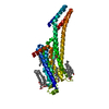 8pwnC 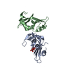 8px0C 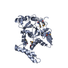 8px1C  8px4C 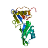 8px5C  8px7C  8px9C 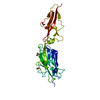 8pxcC  8pxgC  8pxhC  8pxkC 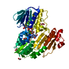 8pxlC 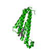 8pyvC 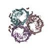 8pyzC  8pz4C 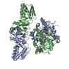 8pz5C C: citing same article ( |
|---|---|
| Similar structure data |
-Links
 PDBj
PDBj- Assembly
| Deposited unit | 
| ||||||||||||
|---|---|---|---|---|---|---|---|---|---|---|---|---|---|
| 1 |
| ||||||||||||
| Unit cell |
| ||||||||||||
| Components on special symmetry positions |
|
-Components
| #1: Protein | Mass: 18652.061 Da / Num. of mol.: 1 Source method: isolated from a genetically manipulated source Source: (gene. exp.) Mammarenavirus whitewaterense / Gene: GPC, GP-C / Production host:  Homo sapiens (human) / References: UniProt: Q911P0 Homo sapiens (human) / References: UniProt: Q911P0 | ||||||||
|---|---|---|---|---|---|---|---|---|---|
| #2: Sugar |   Type: D-saccharide, beta linking / Mass: 221.208 Da / Num. of mol.: 3 / Source method: obtained synthetically / Formula: C8H15NO6 Type: D-saccharide, beta linking / Mass: 221.208 Da / Num. of mol.: 3 / Source method: obtained synthetically / Formula: C8H15NO6#3: Chemical | ChemComp-CD /   Mass: 112.411 Da / Num. of mol.: 6 / Source method: obtained synthetically / Formula: Cd Mass: 112.411 Da / Num. of mol.: 6 / Source method: obtained synthetically / Formula: Cd#4: Chemical |   Mass: 96.063 Da / Num. of mol.: 2 / Source method: obtained synthetically / Formula: SO4 Mass: 96.063 Da / Num. of mol.: 2 / Source method: obtained synthetically / Formula: SO4Has ligand of interest | N | Has protein modification | Y | |
-Experimental details
-Experiment
| Experiment | Method: X-RAY DIFFRACTION / Number of used crystals: 1 |
|---|
- Sample preparation
| Crystal | Density Matthews: 3.31 Å3/Da / Density % sol: 62.79 % |
|---|---|
| Crystal grow | Temperature: 298 K / Method: vapor diffusion, sitting drop Details: 1.0 mg/ml protein, 0.1 M HEPES-Na pH 7.5, and 0.05 M cadmium sulphate |
-Data collection
| Diffraction | Mean temperature: 80 K / Serial crystal experiment: N |
|---|---|
| Diffraction source | Source: SYNCHROTRON / Site: Diamond / Beamline: I23 / Wavelength: 2.7552 Å |
| Detector | Type: DECTRIS PILATUS 12M / Detector: PIXEL / Date: Jul 20, 2016 |
| Radiation | Protocol: SINGLE WAVELENGTH / Monochromatic (M) / Laue (L): M / Scattering type: x-ray |
| Radiation wavelength | Wavelength: 2.7552 Å / Relative weight: 1 |
| Reflection | Resolution: 2.75→46.3 Å / Num. obs: 6783 / % possible obs: 97.2 % / Redundancy: 30.7 % / Biso Wilson estimate: 102.96 Å2 / CC1/2: 0.999 / Rmerge(I) obs: 0.1404 / Rpim(I) all: 0.02522 / Rrim(I) all: 0.1428 / Net I/σ(I): 21.54 |
| Reflection shell | Resolution: 2.75→2.848 Å / Mean I/σ(I) obs: 0.88 / Num. unique obs: 608 / CC1/2: 0.567 |
- Processing
| Software |
| ||||||||||||||||||||||||||||||||||||||||||||||||||||||||||||||||||||||||||||||||||||||||||||||||||||||||||||||||||||||||||||||||||||||||||||||||||||||||||||||||||||||||||||||||||||||
|---|---|---|---|---|---|---|---|---|---|---|---|---|---|---|---|---|---|---|---|---|---|---|---|---|---|---|---|---|---|---|---|---|---|---|---|---|---|---|---|---|---|---|---|---|---|---|---|---|---|---|---|---|---|---|---|---|---|---|---|---|---|---|---|---|---|---|---|---|---|---|---|---|---|---|---|---|---|---|---|---|---|---|---|---|---|---|---|---|---|---|---|---|---|---|---|---|---|---|---|---|---|---|---|---|---|---|---|---|---|---|---|---|---|---|---|---|---|---|---|---|---|---|---|---|---|---|---|---|---|---|---|---|---|---|---|---|---|---|---|---|---|---|---|---|---|---|---|---|---|---|---|---|---|---|---|---|---|---|---|---|---|---|---|---|---|---|---|---|---|---|---|---|---|---|---|---|---|---|---|---|---|---|---|
| Refinement | Method to determine structure: SAD / Resolution: 2.75→46.3 Å / Cor.coef. Fo:Fc: 0.936 / Cor.coef. Fo:Fc free: 0.932 / SU B: 45.082 / SU ML: 0.36 / Cross valid method: THROUGHOUT / ESU R: 0.622 / ESU R Free: 0.339 / Stereochemistry target values: MAXIMUM LIKELIHOOD / Details: HYDROGENS HAVE BEEN ADDED IN THE RIDING POSITIONS
| ||||||||||||||||||||||||||||||||||||||||||||||||||||||||||||||||||||||||||||||||||||||||||||||||||||||||||||||||||||||||||||||||||||||||||||||||||||||||||||||||||||||||||||||||||||||
| Solvent computation | Ion probe radii: 0.8 Å / Shrinkage radii: 0.8 Å / VDW probe radii: 1.2 Å / Solvent model: MASK | ||||||||||||||||||||||||||||||||||||||||||||||||||||||||||||||||||||||||||||||||||||||||||||||||||||||||||||||||||||||||||||||||||||||||||||||||||||||||||||||||||||||||||||||||||||||
| Displacement parameters | Biso mean: 117.52 Å2
| ||||||||||||||||||||||||||||||||||||||||||||||||||||||||||||||||||||||||||||||||||||||||||||||||||||||||||||||||||||||||||||||||||||||||||||||||||||||||||||||||||||||||||||||||||||||
| Refinement step | Cycle: 1 / Resolution: 2.75→46.3 Å
| ||||||||||||||||||||||||||||||||||||||||||||||||||||||||||||||||||||||||||||||||||||||||||||||||||||||||||||||||||||||||||||||||||||||||||||||||||||||||||||||||||||||||||||||||||||||
| Refine LS restraints |
|