 Movie
Movie Controller

[English] 日本語
 Yorodumi
- PDB-8pc0: Sub-tomogram average of the open conformation of the Nap adhesion... -
+ Open data

- Basic information
| Entry | Database: PDB / ID: 8pc0 | ||||||
|---|---|---|---|---|---|---|---|
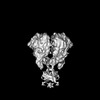
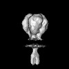
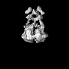
| Title | Sub-tomogram average of the open conformation of the Nap adhesion complex from the human pathogen Mycoplasma genitalium. | ||||||
 Components Components |
| ||||||
 Keywords Keywords | CELL ADHESION / Adhesion / Mycoplasma genitalium | ||||||
| Function / homology |  Function and homology information Function and homology informationadhesion of symbiont to microvasculature / cell adhesion / plasma membrane Similarity search - Function | ||||||
| Biological species |  Mycoplasmoides genitalium G37 (bacteria) Mycoplasmoides genitalium G37 (bacteria) | ||||||
| Method | ELECTRON MICROSCOPY / subtomogram averaging / cryo EM / Resolution: 17 Å | ||||||
 Authors Authors | Sprankel, L. / Scheffer, M.P. / Frangakis, A.F. | ||||||
| Funding support |  Germany, 1items Germany, 1items
| ||||||
 Citation Citation | Journal: PLoS Pathog / Year: 2023 Title: Cryo-electron tomography reveals the binding and release states of the major adhesion complex from Mycoplasma genitalium. Authors: Lasse Sprankel / Margot P Scheffer / Sina Manger / Utz H Ermel / Achilleas S Frangakis / Abstract: The nap particle is an immunogenic surface adhesion complex from Mycoplasma genitalium. It is essential for motility and responsible for binding sialylated oligosaccharides on the surface of the host ...The nap particle is an immunogenic surface adhesion complex from Mycoplasma genitalium. It is essential for motility and responsible for binding sialylated oligosaccharides on the surface of the host cell. The nap particle is composed of two P140-P110 heterodimers, the structure of which was recently solved. However, the interpretation of the mechanism by which the mycoplasma cells orchestrate adhesion remained challenging. Here, we provide cryo-electron tomography structures at ~11 Å resolution, which allow for the distinction between the bound and released state of the nap particle, displaying the in vivo conformational states. Fitting of the atomically resolved structures reveals that bound sialylated oligosaccharides are stabilized by both P110 and P140. Movement of the stalk domains allows for the transfer of conformational changes from the interior of the cell to the binding pocket, thus having the capability of an active release process. It is likely that the same mechanism can be transferred to other Mycoplasma species that belong to the pneumoniae cluster. | ||||||
| History |
|
- Structure visualization
| Structure viewer | Molecule: MolmilJmol/JSmol |
|---|
- Downloads & links
-Download
| PDBx/mmCIF format | 8pc0.cif.gz | 999.1 KB | Display | PDBx/mmCIF format |
|---|---|---|---|---|
| PDB format | pdb8pc0.ent.gz | 828.4 KB | Display | PDB format |
| PDBx/mmJSON format | 8pc0.json.gz | Tree view | PDBx/mmJSON format | |
| Others |  Other downloads Other downloads |
-Validation report
| Arichive directory | https://data.pdbj.org/pub/pdb/validation_reports/pc/8pc0ftp://data.pdbj.org/pub/pdb/validation_reports/pc/8pc0 | HTTPS FTP |
|---|
-Related structure data
| Related structure data | 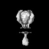 17592MC 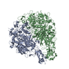 8pbxC 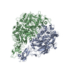 8pbyC 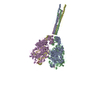 8pbzC 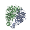 8pc1C C: citing same article ( M: map data used to model this data |
|---|---|
| Similar structure data |


-Links
 PDBj
PDBj- Assembly
| Deposited unit | 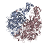
|
|---|---|
| 1 |
|
-Components
-Protein , 2 types, 2 molecules AC
| #1: Protein | Mass: 114553.211 Da / Num. of mol.: 1 / Source method: isolated from a natural source / Source: (natural) Mycoplasmoides genitalium G37 (bacteria) / References: UniProt: P22747 |
|---|---|
| #2: Protein | Mass: 159809.297 Da / Num. of mol.: 1 / Source method: isolated from a natural source / Source: (natural) Mycoplasmoides genitalium G37 (bacteria) / References: UniProt: P20796 |
-Sugars , 1 types, 1 molecules
| #3: Polysaccharide | N-acetyl-alpha-neuraminic acid-(2-6)-beta-D-galactopyranose-(1-4)-1,5-anhydro-D-glucitol Type: oligosaccharide / Mass: 617.552 Da / Num. of mol.: 1 Source method: isolated from a genetically manipulated source |
|---|
-Non-polymers , 3 types, 101 molecules 


| #4: Chemical | ChemComp-K /  Mass: 39.098 Da / Num. of mol.: 1 / Source method: obtained synthetically / Formula: K Mass: 39.098 Da / Num. of mol.: 1 / Source method: obtained synthetically / Formula: K |
|---|---|
| #5: Chemical | ChemComp-PO4 /  Mass: 94.971 Da / Num. of mol.: 1 / Source method: obtained synthetically / Formula: PO4 Mass: 94.971 Da / Num. of mol.: 1 / Source method: obtained synthetically / Formula: PO4 |
| #6: Water | ChemComp-HOH / Mass: 18.015 Da / Num. of mol.: 99 / Source method: isolated from a natural source / Formula: H2O |
-Details
| Has ligand of interest | N |
|---|
-Experimental details
-Experiment
| Experiment | Method: ELECTRON MICROSCOPY |
|---|---|
| EM experiment | Aggregation state: CELL / 3D reconstruction method: subtomogram averaging |
- Sample preparation
| Component | Name: Mildly lysed M. genitalium cells / Type: CELL / Entity ID: #1-#2 / Source: NATURAL |
|---|---|
| Source (natural) | Organism: Mycoplasmoides genitalium G37 (bacteria) |
| Buffer solution | pH: 7.5 |
| Specimen | Embedding applied: NO / Shadowing applied: NO / Staining applied: NO / Vitrification applied: YES / Details: Mildy lysed M. genitalium cells |
| Vitrification | Instrument: FEI VITROBOT MARK IV / Cryogen name: ETHANE / Humidity: 100 % / Chamber temperature: 277.15 K |
- Electron microscopy imaging
| Experimental equipment |  Model: Titan Krios / Image courtesy: FEI Company |
|---|---|
| Microscopy | Model: FEI TITAN KRIOS |
| Electron gun | Electron source:  FIELD EMISSION GUN / Accelerating voltage: 300 kV / Illumination mode: FLOOD BEAM FIELD EMISSION GUN / Accelerating voltage: 300 kV / Illumination mode: FLOOD BEAM |
| Electron lens | Mode: BRIGHT FIELD / Nominal magnification: 105000 X / Nominal defocus max: 4000 nm / Nominal defocus min: 2000 nm / Cs: 2.7 mm / C2 aperture diameter: 70 µm / Alignment procedure: BASIC |
| Specimen holder | Cryogen: NITROGEN / Specimen holder model: FEI TITAN KRIOS AUTOGRID HOLDER |
| Image recording | Electron dose: 3 e/Å2 / Avg electron dose per subtomogram: 120 e/Å2 / Detector mode: SUPER-RESOLUTION / Film or detector model: GATAN K2 SUMMIT (4k x 4k) |
- Processing
| CTF correction | Type: PHASE FLIPPING AND AMPLITUDE CORRECTION |
|---|---|
| Symmetry | Point symmetry: C1 (asymmetric) |
| 3D reconstruction | Resolution: 17 Å / Resolution method: FSC 0.143 CUT-OFF / Num. of particles: 9689 / Symmetry type: POINT |
| EM volume selection | Num. of tomograms: 420 / Num. of volumes extracted: 36720 |
