Movie
Movie Controller

[English] 日本語
 Yorodumi
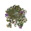
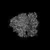
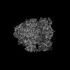
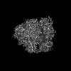
- PDB-8p6p: Mycoplasma pneumoniae small ribosomal subunit in chloramphenicol-... -
+ Open data

- Basic information
| Entry | Database: PDB / ID: 8p6p | |||||||||
|---|---|---|---|---|---|---|---|---|---|---|
| Title | Mycoplasma pneumoniae small ribosomal subunit in chloramphenicol-treated cells | |||||||||
 Components Components |
| |||||||||
 Keywords Keywords | TRANSLATION / In situ / Ribosome / Chloramphenicol / cryo-ET | |||||||||
| Function / homology |  Function and homology information Function and homology informationribosomal small subunit assembly / ribosomal small subunit biogenesis / small ribosomal subunit / small ribosomal subunit rRNA binding / cytosolic small ribosomal subunit / tRNA binding / rRNA binding / structural constituent of ribosome / ribosome / translation ...ribosomal small subunit assembly / ribosomal small subunit biogenesis / small ribosomal subunit / small ribosomal subunit rRNA binding / cytosolic small ribosomal subunit / tRNA binding / rRNA binding / structural constituent of ribosome / ribosome / translation / ribonucleoprotein complex / mRNA binding / RNA binding / zinc ion binding / cytosol / cytoplasm Similarity search - Function | |||||||||
| Biological species |  Mycoplasmoides pneumoniae M129 (bacteria) Mycoplasmoides pneumoniae M129 (bacteria) | |||||||||
| Method | ELECTRON MICROSCOPY / subtomogram averaging / cryo EM / Resolution: 3.2 Å | |||||||||
 Authors Authors | Schacherl, M. / Xue, L. / Spahn, C.M.T. / Mahamid, J. | |||||||||
| Funding support |  United States, United States,  Germany, 2items Germany, 2items
| |||||||||
 Citation Citation | Journal: Nat Struct Mol Biol / Year: 2025 Title: Structural insights into context-dependent inhibitory mechanisms of chloramphenicol in cells. Authors: Liang Xue / Christian M T Spahn / Magdalena Schacherl / Julia Mahamid /  Abstract: Ribosome-targeting antibiotics represent an important class of antimicrobial drugs. Chloramphenicol (Cm) is a well-studied ribosomal peptidyl transferase center (PTC) binder and growing evidence ...Ribosome-targeting antibiotics represent an important class of antimicrobial drugs. Chloramphenicol (Cm) is a well-studied ribosomal peptidyl transferase center (PTC) binder and growing evidence suggests that its inhibitory action depends on the sequence of the nascent peptide. How such selective inhibition on the molecular scale manifests on the cellular level remains unclear. Here, we use cryo-electron tomography to analyze the impact of Cm inside the bacterium Mycoplasma pneumoniae. By resolving the Cm-bound ribosomes to 3.0 Å, we elucidate Cm's coordination with natural nascent peptides and transfer RNAs in the PTC. We find that Cm leads to the accumulation of a number of translation elongation states, indicating ongoing futile accommodation cycles, and to extensive ribosome collisions. We, thus, suggest that, beyond its direct inhibition of protein synthesis, the action of Cm may involve the activation of cellular stress responses. This work exemplifies how in-cell structural biology can expand the understanding of mechanisms of action for extensively studied antibiotics. | |||||||||
| History |
|
- Structure visualization
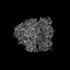
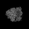
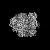
| Structure viewer | Molecule: MolmilJmol/JSmol |
|---|
- Downloads & links
-Download
| PDBx/mmCIF format | 8p6p.cif.gz | 1.3 MB | Display | PDBx/mmCIF format |
|---|---|---|---|---|
| PDB format | pdb8p6p.ent.gz | 982.3 KB | Display | PDB format |
| PDBx/mmJSON format | 8p6p.json.gz | Tree view | PDBx/mmJSON format | |
| Others |  Other downloads Other downloads |
-Validation report
| Arichive directory | https://data.pdbj.org/pub/pdb/validation_reports/p6/8p6pftp://data.pdbj.org/pub/pdb/validation_reports/p6/8p6p | HTTPS FTP |
|---|
-Related structure data
| Related structure data |  17133MC  8p7xC  8p7yC  8p8bC  8p8vC  8p8wC M: map data used to model this data C: citing same article ( |
|---|---|
| Similar structure data | |
| Experimental dataset #1 | Data reference: 10.6019/EMPIAR-11520 / Data set type: EMPIAR |


-Links
 PDBj
PDBj


- Assembly
| Deposited unit | 
|
|---|---|
| 1 |
|
-Components
-RNA chain , 5 types, 5 molecules 3578Y
| #1: RNA chain | Mass: 940911.500 Da / Num. of mol.: 1 / Source method: isolated from a natural source / Source: (natural) Mycoplasmoides pneumoniae M129 (bacteria) |
|---|---|
| #2: RNA chain | Mass: 491326.844 Da / Num. of mol.: 1 / Source method: isolated from a natural source / Source: (natural) Mycoplasmoides pneumoniae M129 (bacteria) / References: GenBank: 26117688 |
| #3: RNA chain | Mass: 24136.244 Da / Num. of mol.: 1 / Source method: isolated from a natural source / Source: (natural) Mycoplasmoides pneumoniae M129 (bacteria) / References: GenBank: 26117688 |
| #4: RNA chain | Mass: 24401.449 Da / Num. of mol.: 1 / Source method: isolated from a natural source / Source: (natural) Mycoplasmoides pneumoniae M129 (bacteria) / References: GenBank: 26117688 |
| #25: RNA chain | Mass: 6698.029 Da / Num. of mol.: 1 / Source method: isolated from a natural source / Source: (natural) Mycoplasmoides pneumoniae M129 (bacteria) |
-30S ribosomal protein ... , 20 types, 20 molecules ABCDEFGHIJKLMNOPQRST
| #5: Protein | Mass: 33468.629 Da / Num. of mol.: 1 / Source method: isolated from a natural source / Source: (natural) Mycoplasmoides pneumoniae M129 (bacteria) / References: UniProt: P75560 |
|---|---|
| #6: Protein | Mass: 30657.205 Da / Num. of mol.: 1 / Source method: isolated from a natural source / Source: (natural) Mycoplasmoides pneumoniae M129 (bacteria) / References: UniProt: P41205 |
| #7: Protein | Mass: 23817.645 Da / Num. of mol.: 1 / Source method: isolated from a natural source / Source: (natural) Mycoplasmoides pneumoniae M129 (bacteria) / References: UniProt: P46775 |
| #8: Protein | Mass: 24138.008 Da / Num. of mol.: 1 / Source method: isolated from a natural source / Source: (natural) Mycoplasmoides pneumoniae M129 (bacteria) / References: UniProt: Q50301 |
| #9: Protein | Mass: 25430.465 Da / Num. of mol.: 1 / Source method: isolated from a natural source / Source: (natural) Mycoplasmoides pneumoniae M129 (bacteria) / References: UniProt: P75543 |
| #10: Protein | Mass: 17897.029 Da / Num. of mol.: 1 / Source method: isolated from a natural source / Source: (natural) Mycoplasmoides pneumoniae M129 (bacteria) / References: UniProt: P75545 |
| #11: Protein | Mass: 15903.083 Da / Num. of mol.: 1 / Source method: isolated from a natural source / Source: (natural) Mycoplasmoides pneumoniae M129 (bacteria) / References: UniProt: Q50304 |
| #12: Protein | Mass: 15149.735 Da / Num. of mol.: 1 / Source method: isolated from a natural source / Source: (natural) Mycoplasmoides pneumoniae M129 (bacteria) / References: UniProt: P75179 |
| #13: Protein | Mass: 12226.571 Da / Num. of mol.: 1 / Source method: isolated from a natural source / Source: (natural) Mycoplasmoides pneumoniae M129 (bacteria) / References: UniProt: P75581 |
| #14: Protein | Mass: 12709.851 Da / Num. of mol.: 1 / Source method: isolated from a natural source / Source: (natural) Mycoplasmoides pneumoniae M129 (bacteria) / References: UniProt: Q50296 |
| #15: Protein | Mass: 15666.580 Da / Num. of mol.: 1 / Source method: isolated from a natural source / Source: (natural) Mycoplasmoides pneumoniae M129 (bacteria) / References: UniProt: P75546 |
| #16: Protein | Mass: 14209.607 Da / Num. of mol.: 1 / Source method: isolated from a natural source / Source: (natural) Mycoplasmoides pneumoniae M129 (bacteria) / References: UniProt: Q50297 |
| #17: Protein | Mass: 6901.364 Da / Num. of mol.: 1 / Source method: isolated from a natural source / Source: (natural) Mycoplasmoides pneumoniae M129 (bacteria) / References: UniProt: Q50305 |
| #18: Protein | Mass: 9921.687 Da / Num. of mol.: 1 / Source method: isolated from a natural source / Source: (natural) Mycoplasmoides pneumoniae M129 (bacteria) / References: UniProt: P75173 |
| #19: Protein | Mass: 10806.921 Da / Num. of mol.: 1 / Source method: isolated from a natural source / Source: (natural) Mycoplasmoides pneumoniae M129 (bacteria) / References: UniProt: A0A0H3DLS7 |
| #20: Protein | Mass: 9859.643 Da / Num. of mol.: 1 / Source method: isolated from a natural source / Source: (natural) Mycoplasmoides pneumoniae M129 (bacteria) / References: UniProt: Q50309 |
| #21: Protein | Mass: 12411.522 Da / Num. of mol.: 1 / Source method: isolated from a natural source / Source: (natural) Mycoplasmoides pneumoniae M129 (bacteria) / References: UniProt: P75541 |
| #22: Protein | Mass: 10057.626 Da / Num. of mol.: 1 / Source method: isolated from a natural source / Source: (natural) Mycoplasmoides pneumoniae M129 (bacteria) / References: UniProt: P75576 |
| #23: Protein | Mass: 9993.599 Da / Num. of mol.: 1 / Source method: isolated from a natural source / Source: (natural) Mycoplasmoides pneumoniae M129 (bacteria) / References: UniProt: P75237 |
| #24: Protein | Mass: 7539.182 Da / Num. of mol.: 1 / Source method: isolated from a natural source / Source: (natural) Mycoplasmoides pneumoniae M129 (bacteria) / References: UniProt: P57079 |
-Protein , 1 types, 1 molecules x
| #26: Protein | Mass: 10980.657 Da / Num. of mol.: 1 / Source method: isolated from a natural source / Source: (natural) Mycoplasmoides pneumoniae M129 (bacteria) / References: UniProt: P78020 |
|---|
-Non-polymers , 6 types, 112 molecules 
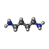


| #27: Chemical |  Mass: 145.246 Da / Num. of mol.: 2 / Source method: obtained synthetically / Formula: C7H19N3 Mass: 145.246 Da / Num. of mol.: 2 / Source method: obtained synthetically / Formula: C7H19N3#28: Chemical |  Mass: 88.151 Da / Num. of mol.: 2 / Source method: obtained synthetically / Formula: C4H12N2 Mass: 88.151 Da / Num. of mol.: 2 / Source method: obtained synthetically / Formula: C4H12N2#29: Chemical | ChemComp-N2P / |  Mass: 102.178 Da / Num. of mol.: 1 / Source method: obtained synthetically / Formula: C5H14N2 Mass: 102.178 Da / Num. of mol.: 1 / Source method: obtained synthetically / Formula: C5H14N2#30: Chemical | ChemComp-MG /  Mass: 24.305 Da / Num. of mol.: 103 / Source method: obtained synthetically / Formula: Mg Mass: 24.305 Da / Num. of mol.: 103 / Source method: obtained synthetically / Formula: Mg#31: Chemical |  Mass: 65.409 Da / Num. of mol.: 2 / Source method: obtained synthetically / Formula: Zn Mass: 65.409 Da / Num. of mol.: 2 / Source method: obtained synthetically / Formula: Zn#32: Water | ChemComp-HOH / | Mass: 18.015 Da / Num. of mol.: 2 / Source method: isolated from a natural source / Formula: H2O |
|---|
-Details
| Has ligand of interest | N |
|---|---|
| Has protein modification | N |
-Experimental details
-Experiment
| Experiment | Method: ELECTRON MICROSCOPY |
|---|---|
| EM experiment | Aggregation state: CELL / 3D reconstruction method: subtomogram averaging |
- Sample preparation
| Component | Name: Mycoplasma pneumoniae M129 cells treated with chloramphenicol Type: CELL Details: Small ribosomal subunit with some elements of the large ribosomal subunit. Focused refinement on 30S subunit of the in situ 70S subunit map. Entity ID: #1-#26 / Source: NATURAL |
|---|---|
| Source (natural) | Organism: Mycoplasmoides pneumoniae M129 (bacteria) / Cellular location: Cytoplasm |
| Buffer solution | pH: 7.4 Details: The modified Hayflick medium: 14.7g/L Difco PPLO(Becton Dickinson), 20% (v/v) Gibco horse serum(New Zealand origin), 100 mM HEPES-Na; pH 7.4, 1% (w/w) glucose, 0.002% (w/w) phenol red, 1000 U/mL penicillin G. |
| Specimen | Embedding applied: NO / Shadowing applied: NO / Staining applied: NO / Vitrification applied: YES Details: Mycoplasma pneumoniae M129 cells were grown on gold Quantifoil grids at 37 Celsius in the modified Hayflick medium. Treatment with chloramphenicol at the final concentration of 0.2 mg/ml was ...Details: Mycoplasma pneumoniae M129 cells were grown on gold Quantifoil grids at 37 Celsius in the modified Hayflick medium. Treatment with chloramphenicol at the final concentration of 0.2 mg/ml was performed for about 15 minutes before plunge freezing. |
| Specimen support | Grid material: GOLD / Grid mesh size: 200 divisions/in. / Grid type: Quantifoil R2/1 |
| Vitrification | Instrument: HOMEMADE PLUNGER / Cryogen name: ETHANE-PROPANE Details: Back-side blotting for 2-3 seconds before plunging using a manual plunger without an environmental control chamber. |
- Electron microscopy imaging
| Experimental equipment |  Model: Titan Krios / Image courtesy: FEI Company |
|---|---|
| Microscopy | Model: FEI TITAN KRIOS |
| Electron gun | Electron source:  FIELD EMISSION GUN / Accelerating voltage: 300 kV / Illumination mode: FLOOD BEAM FIELD EMISSION GUN / Accelerating voltage: 300 kV / Illumination mode: FLOOD BEAM |
| Electron lens | Mode: BRIGHT FIELD / Nominal magnification: 64000 X / Nominal defocus max: 3250 nm / Nominal defocus min: 1000 nm / Cs: 2.7 mm / Alignment procedure: COMA FREE |
| Specimen holder | Cryogen: NITROGEN / Specimen holder model: FEI TITAN KRIOS AUTOGRID HOLDER |
| Image recording | Electron dose: 3.34 e/Å2 / Avg electron dose per subtomogram: 137 e/Å2 / Film or detector model: GATAN K3 BIOQUANTUM (6k x 4k) / Num. of real images: 1 Details: Gatan K3 camera in non-CDS counting mode, targeted dose rate on camera ~20 e/pixel/second, 10 frames per tilt image, constant exposure time for each tilt, pixel size 1.329A |
| EM imaging optics | Energyfilter name: GIF Bioquantum / Energyfilter slit width: 20 eV |
- Processing
| EM software |
| |||||||||||||||||||||||||||||||||||||||||||||||||||||||
|---|---|---|---|---|---|---|---|---|---|---|---|---|---|---|---|---|---|---|---|---|---|---|---|---|---|---|---|---|---|---|---|---|---|---|---|---|---|---|---|---|---|---|---|---|---|---|---|---|---|---|---|---|---|---|---|---|
| CTF correction | Details: CTF estimation and 3D CTF correction are done in Warp Type: PHASE FLIPPING AND AMPLITUDE CORRECTION | |||||||||||||||||||||||||||||||||||||||||||||||||||||||
| Symmetry | Point symmetry: C1 (asymmetric) | |||||||||||||||||||||||||||||||||||||||||||||||||||||||
| 3D reconstruction | Resolution: 3.2 Å / Resolution method: FSC 0.143 CUT-OFF / Num. of particles: 30774 / Algorithm: FOURIER SPACE / Details: half maps from Warp/M / Symmetry type: POINT | |||||||||||||||||||||||||||||||||||||||||||||||||||||||
| EM volume selection | Num. of tomograms: 139 / Num. of volumes extracted: 30774 | |||||||||||||||||||||||||||||||||||||||||||||||||||||||
| Atomic model building | Protocol: RIGID BODY FIT / Space: REAL / Target criteria: Cross-correlation coefficient | |||||||||||||||||||||||||||||||||||||||||||||||||||||||
| Atomic model building |
| |||||||||||||||||||||||||||||||||||||||||||||||||||||||
| Refine LS restraints |
|