Movie
Movie Controller

[English] 日本語
 Yorodumi
- PDB-7ooc: Mycoplasma pneumoniae 30S subunit of ribosomes in chloramphenicol... -
+ Open data

- Basic information
| Entry | Database: PDB / ID: 7ooc | ||||||||||||
|---|---|---|---|---|---|---|---|---|---|---|---|---|---|
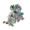
| Title | Mycoplasma pneumoniae 30S subunit of ribosomes in chloramphenicol-treated cells | ||||||||||||
 Components Components |
| ||||||||||||
 Keywords Keywords | RIBOSOME / In-cell cryo-electron tomography chloramphenicol-treated sub-tomogram analysis | ||||||||||||
| Function / homology |  Function and homology information Function and homology informationribosomal small subunit assembly / ribosomal small subunit biogenesis / small ribosomal subunit / small ribosomal subunit rRNA binding / cytosolic small ribosomal subunit / tRNA binding / rRNA binding / structural constituent of ribosome / ribosome / translation ...ribosomal small subunit assembly / ribosomal small subunit biogenesis / small ribosomal subunit / small ribosomal subunit rRNA binding / cytosolic small ribosomal subunit / tRNA binding / rRNA binding / structural constituent of ribosome / ribosome / translation / ribonucleoprotein complex / mRNA binding / RNA binding / zinc ion binding / cytoplasm / cytosol Similarity search - Function | ||||||||||||
| Biological species |  Mycoplasma pneumoniae (Filterable agent of primary atypical pneumonia) Mycoplasma pneumoniae (Filterable agent of primary atypical pneumonia) | ||||||||||||
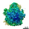
| Method | ELECTRON MICROSCOPY / subtomogram averaging / cryo EM / Resolution: 3.7 Å | ||||||||||||
 Authors Authors | Xue, L. / Lenz, S. / Rappsilber, J. / Mahamid, J. | ||||||||||||
| Funding support |  Germany, Germany,  United Kingdom, 3items United Kingdom, 3items
| ||||||||||||
 Citation Citation | Journal: Nature / Year: 2022 Title: Visualizing translation dynamics at atomic detail inside a bacterial cell. Authors: Liang Xue / Swantje Lenz / Maria Zimmermann-Kogadeeva / Dimitry Tegunov / Patrick Cramer / Peer Bork / Juri Rappsilber / Julia Mahamid /  Abstract: Translation is the fundamental process of protein synthesis and is catalysed by the ribosome in all living cells. Here we use advances in cryo-electron tomography and sub-tomogram analysis to ...Translation is the fundamental process of protein synthesis and is catalysed by the ribosome in all living cells. Here we use advances in cryo-electron tomography and sub-tomogram analysis to visualize the structural dynamics of translation inside the bacterium Mycoplasma pneumoniae. To interpret the functional states in detail, we first obtain a high-resolution in-cell average map of all translating ribosomes and build an atomic model for the M. pneumoniae ribosome that reveals distinct extensions of ribosomal proteins. Classification then resolves 13 ribosome states that differ in their conformation and composition. These recapitulate major states that were previously resolved in vitro, and reflect intermediates during active translation. On the basis of these states, we animate translation elongation inside native cells and show how antibiotics reshape the cellular translation landscapes. During translation elongation, ribosomes often assemble in defined three-dimensional arrangements to form polysomes. By mapping the intracellular organization of translating ribosomes, we show that their association into polysomes involves a local coordination mechanism that is mediated by the ribosomal protein L9. We propose that an extended conformation of L9 within polysomes mitigates collisions to facilitate translation fidelity. Our work thus demonstrates the feasibility of visualizing molecular processes at atomic detail inside cells. #1: Journal: Nature / Year: 2022Title: Visualizing translation dynamics at atomic detail inside a bacterial cell. Authors: Liang Xue / Swantje Lenz / Maria Zimmermann-Kogadeeva / Dimitry Tegunov / Patrick Cramer / Peer Bork / Juri Rappsilber / Julia Mahamid / Abstract: Translation is the fundamental process of protein synthesis and is catalysed by the ribosome in all living cells. Here we use advances in cryo-electron tomography and sub-tomogram analysis to ...Translation is the fundamental process of protein synthesis and is catalysed by the ribosome in all living cells. Here we use advances in cryo-electron tomography and sub-tomogram analysis to visualize the structural dynamics of translation inside the bacterium Mycoplasma pneumoniae. To interpret the functional states in detail, we first obtain a high-resolution in-cell average map of all translating ribosomes and build an atomic model for the M. pneumoniae ribosome that reveals distinct extensions of ribosomal proteins. Classification then resolves 13 ribosome states that differ in their conformation and composition. These recapitulate major states that were previously resolved in vitro, and reflect intermediates during active translation. On the basis of these states, we animate translation elongation inside native cells and show how antibiotics reshape the cellular translation landscapes. During translation elongation, ribosomes often assemble in defined three-dimensional arrangements to form polysomes. By mapping the intracellular organization of translating ribosomes, we show that their association into polysomes involves a local coordination mechanism that is mediated by the ribosomal protein L9. We propose that an extended conformation of L9 within polysomes mitigates collisions to facilitate translation fidelity. Our work thus demonstrates the feasibility of visualizing molecular processes at atomic detail inside cells. #2: Journal: Biorxiv / Year: 2021Title: Visualizing translation dynamics at atomic detail inside a bacterial cell Authors: Xue, L. / Lenz, S. / Zimmermann-Kogadeeva, M. / Tegunov, D. / Cramer, P. / Bork, P. / Rappsilber, J. / Mahamid, J. | ||||||||||||
| History |
|
- Structure visualization
| Structure viewer | Molecule: MolmilJmol/JSmol |
|---|
- Downloads & links
-Download
| PDBx/mmCIF format | 7ooc.cif.gz | 1.1 MB | Display | PDBx/mmCIF format |
|---|---|---|---|---|
| PDB format | pdb7ooc.ent.gz | 890.7 KB | Display | PDB format |
| PDBx/mmJSON format | 7ooc.json.gz | Tree view | PDBx/mmJSON format | |
| Others |  Other downloads Other downloads |
-Validation report
| Arichive directory | https://data.pdbj.org/pub/pdb/validation_reports/oo/7oocftp://data.pdbj.org/pub/pdb/validation_reports/oo/7ooc | HTTPS FTP |
|---|
-Related structure data
| Related structure data | 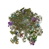 7oodC 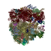 7p6zC 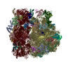 7pahC 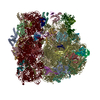 7paiC 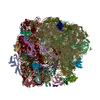 7pajC 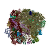 7pakC 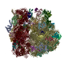 7palC 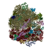 7pamC 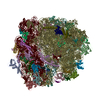 7panC 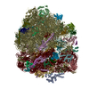 7paoC 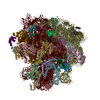 7paqC 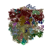 7parC 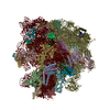 7pasC 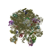 7patC  7pauC 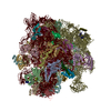 7ph9C 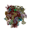 7phaC 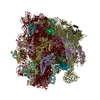 7phbC 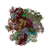 7phcC 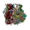 7pi8C 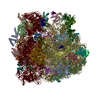 7pi9C 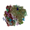 7piaC 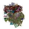 7pibC 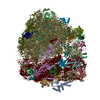 7picC 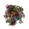 7pioC 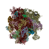 7pipC 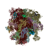 7piqC 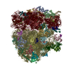 7pirC 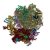 7pisC 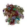 7pitC M: map data used to model this data C: citing same article ( |
|---|---|
| Similar structure data | |
| EM raw data | EMPIAR-10499 (Title: Tilt series of native M. pneumoniae cells treated with chloramphenicol Data size: 83.8 Data #1: Unaligned tilt movies of M. pneumoniae [tilt series]) |
| Experimental dataset #1 | Data reference: 10.6019/EMPIAR-10499 / Data set type: EMPIAR |


-Links
 PDBj
PDBj


- Assembly
| Deposited unit | 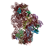
|
|---|---|
| 1 |
|
-Components
-30S ribosomal protein ... , 20 types, 20 molecules BDFAHJCSOKMILNRTGQEP
| #1: Protein | Mass: 30657.205 Da / Num. of mol.: 1 / Source method: isolated from a natural source Source: (natural) Mycoplasma pneumoniae (strain ATCC 29342 / M129) (bacteria)Strain: ATCC 29342 / M129 / References: UniProt: P41205 |
|---|---|
| #2: Protein | Mass: 24138.008 Da / Num. of mol.: 1 / Source method: isolated from a natural source Source: (natural) Mycoplasma pneumoniae (strain ATCC 29342 / M129) (bacteria)Strain: ATCC 29342 / M129 / References: UniProt: Q50301 |
| #3: Protein | Mass: 17897.029 Da / Num. of mol.: 1 / Source method: isolated from a natural source Source: (natural) Mycoplasma pneumoniae (strain ATCC 29342 / M129) (bacteria)Strain: ATCC 29342 / M129 / References: UniProt: P75545 |
| #4: Protein | Mass: 33468.629 Da / Num. of mol.: 1 / Source method: isolated from a natural source Source: (natural) Mycoplasma pneumoniae (strain ATCC 29342 / M129) (bacteria)Strain: ATCC 29342 / M129 / References: UniProt: P75560 |
| #5: Protein | Mass: 15149.735 Da / Num. of mol.: 1 / Source method: isolated from a natural source Source: (natural) Mycoplasma pneumoniae (strain ATCC 29342 / M129) (bacteria)Strain: ATCC 29342 / M129 / References: UniProt: P75179 |
| #6: Protein | Mass: 12709.851 Da / Num. of mol.: 1 / Source method: isolated from a natural source Source: (natural) Mycoplasma pneumoniae (strain ATCC 29342 / M129) (bacteria)Strain: ATCC 29342 / M129 / References: UniProt: Q50296 |
| #7: Protein | Mass: 23817.645 Da / Num. of mol.: 1 / Source method: isolated from a natural source Source: (natural) Mycoplasma pneumoniae (strain ATCC 29342 / M129) (bacteria)Strain: ATCC 29342 / M129 / References: UniProt: P46775 |
| #8: Protein | Mass: 9993.599 Da / Num. of mol.: 1 / Source method: isolated from a natural source Source: (natural) Mycoplasma pneumoniae (strain ATCC 29342 / M129) (bacteria)Strain: ATCC 29342 / M129 / References: UniProt: P75237 |
| #9: Protein | Mass: 10806.921 Da / Num. of mol.: 1 / Source method: isolated from a natural source Source: (natural) Mycoplasma pneumoniae (strain ATCC 29342 / M129) (bacteria)Strain: ATCC 29342 / M129 / References: UniProt: A0A0H3DLS7 |
| #10: Protein | Mass: 15666.580 Da / Num. of mol.: 1 / Source method: isolated from a natural source Source: (natural) Mycoplasma pneumoniae (strain ATCC 29342 / M129) (bacteria)Strain: ATCC 29342 / M129 / References: UniProt: P75546 |
| #11: Protein | Mass: 6901.364 Da / Num. of mol.: 1 / Source method: isolated from a natural source Source: (natural) Mycoplasma pneumoniae (strain ATCC 29342 / M129) (bacteria)Strain: ATCC 29342 / M129 / References: UniProt: Q50305 |
| #12: Protein | Mass: 12226.571 Da / Num. of mol.: 1 / Source method: isolated from a natural source Source: (natural) Mycoplasma pneumoniae (strain ATCC 29342 / M129) (bacteria)Strain: ATCC 29342 / M129 / References: UniProt: P75581 |
| #13: Protein | Mass: 14209.607 Da / Num. of mol.: 1 / Source method: isolated from a natural source Source: (natural) Mycoplasma pneumoniae (strain ATCC 29342 / M129) (bacteria)Strain: ATCC 29342 / M129 / References: UniProt: Q50297 |
| #14: Protein | Mass: 9921.687 Da / Num. of mol.: 1 / Source method: isolated from a natural source Source: (natural) Mycoplasma pneumoniae (strain ATCC 29342 / M129) (bacteria)Strain: ATCC 29342 / M129 / References: UniProt: P75173 |
| #15: Protein | Mass: 10057.626 Da / Num. of mol.: 1 / Source method: isolated from a natural source Source: (natural) Mycoplasma pneumoniae (strain ATCC 29342 / M129) (bacteria)Strain: ATCC 29342 / M129 / References: UniProt: P75576 |
| #16: Protein | Mass: 7539.182 Da / Num. of mol.: 1 / Source method: isolated from a natural source Source: (natural) Mycoplasma pneumoniae (strain ATCC 29342 / M129) (bacteria)Strain: ATCC 29342 / M129 / References: UniProt: P57079 |
| #17: Protein | Mass: 15903.083 Da / Num. of mol.: 1 / Source method: isolated from a natural source Source: (natural) Mycoplasma pneumoniae (strain ATCC 29342 / M129) (bacteria)Strain: ATCC 29342 / M129 / References: UniProt: Q50304 |
| #18: Protein | Mass: 12411.522 Da / Num. of mol.: 1 / Source method: isolated from a natural source Source: (natural) Mycoplasma pneumoniae (strain ATCC 29342 / M129) (bacteria)Strain: ATCC 29342 / M129 / References: UniProt: P75541 |
| #19: Protein | Mass: 25430.465 Da / Num. of mol.: 1 / Source method: isolated from a natural source Source: (natural) Mycoplasma pneumoniae (strain ATCC 29342 / M129) (bacteria)Strain: ATCC 29342 / M129 / References: UniProt: P75543 |
| #20: Protein | Mass: 9859.643 Da / Num. of mol.: 1 / Source method: isolated from a natural source Source: (natural) Mycoplasma pneumoniae (strain ATCC 29342 / M129) (bacteria)Strain: ATCC 29342 / M129 / References: UniProt: Q50309 |
-RNA chain / Non-polymers , 2 types, 3 molecules 5
| #21: RNA chain | Mass: 491243.625 Da / Num. of mol.: 1 / Source method: isolated from a natural source Source: (natural) Mycoplasma pneumoniae (strain ATCC 29342 / M129) (bacteria)Strain: ATCC 29342 / M129 |
|---|---|
| #22: Chemical |  Mass: 65.409 Da / Num. of mol.: 2 / Source method: obtained synthetically / Formula: Zn Mass: 65.409 Da / Num. of mol.: 2 / Source method: obtained synthetically / Formula: Zn |
-Details
| Has ligand of interest | N |
|---|
-Experimental details
-Experiment
| Experiment | Method: ELECTRON MICROSCOPY |
|---|---|
| EM experiment | Aggregation state: CELL / 3D reconstruction method: subtomogram averaging |
- Sample preparation
| Component | Name: cryo-electron tomograms of chloramphenicol-treated Mycoplasma pneumoniae cells Type: RIBOSOME Details: ribosome sub-tomograms extracted in silico from cell tomograms, focused refinement on 30S Entity ID: #1-#21 / Source: NATURAL |
|---|---|
| Source (natural) | Organism: Mycoplasma pneumoniae M129 (bacteria) |
| Buffer solution | pH: 7.4 |
| Specimen | Embedding applied: NO / Shadowing applied: NO / Staining applied: NO / Vitrification applied: YES Details: Mycoplasma pneumoniae M129 cells grown on gold Quantifoil grids at 37 degrees Celsius before plunge freezing. Quick wash with PBS solution containing gold fiducial beads before blotting. |
| Specimen support | Grid material: GOLD / Grid type: Quantifoil R2/1 |
| Vitrification | Instrument: HOMEMADE PLUNGER / Cryogen name: ETHANE-PROPANE Details: back-side blotting for 2-3 second before plunging manual plunger without an environmental chamber |
- Electron microscopy imaging
| Experimental equipment |  Model: Titan Krios / Image courtesy: FEI Company |
|---|---|
| Microscopy | Model: FEI TITAN KRIOS |
| Electron gun | Electron source:  FIELD EMISSION GUN / Accelerating voltage: 300 kV / Illumination mode: FLOOD BEAM FIELD EMISSION GUN / Accelerating voltage: 300 kV / Illumination mode: FLOOD BEAM |
| Electron lens | Mode: BRIGHT FIELD / Nominal magnification: 81000 X / Nominal defocus max: 3750 nm / Nominal defocus min: 1500 nm / Cs: 2.7 mm |
| Specimen holder | Cryogen: NITROGEN / Specimen holder model: FEI TITAN KRIOS AUTOGRID HOLDER |
| Image recording | Electron dose: 3.2 e/Å2 / Detector mode: COUNTING / Film or detector model: GATAN K2 SUMMIT (4k x 4k) |
- Processing
| EM software |
| ||||||||||||||||||||
|---|---|---|---|---|---|---|---|---|---|---|---|---|---|---|---|---|---|---|---|---|---|
| CTF correction | Type: PHASE FLIPPING AND AMPLITUDE CORRECTION | ||||||||||||||||||||
| Symmetry | Point symmetry: C1 (asymmetric) | ||||||||||||||||||||
| 3D reconstruction | Resolution: 3.7 Å / Resolution method: FSC 0.143 CUT-OFF / Num. of particles: 17890 Details: Map generated by focused refinement on the 30S subunit in M Symmetry type: POINT | ||||||||||||||||||||
| EM volume selection | Num. of tomograms: 65 / Num. of volumes extracted: 17890 | ||||||||||||||||||||
| Atomic model building | Protocol: AB INITIO MODEL / Space: REAL | ||||||||||||||||||||
| Atomic model building | 3D fitting-ID: 1 / Source name: PDB / Type: experimental model
|