Movie
Movie Controller

[English] 日本語
 Yorodumi
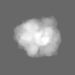
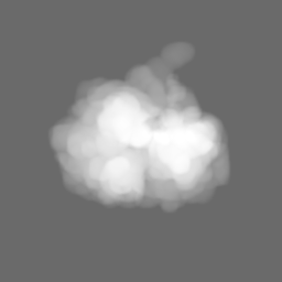
- EMDB-17141: 70S ribosome with EF-Tu-tRNA, P and E-site tRNAs in chloramphenic... -
+ Open data

- Basic information
| Entry |  | |||||||||
|---|---|---|---|---|---|---|---|---|---|---|
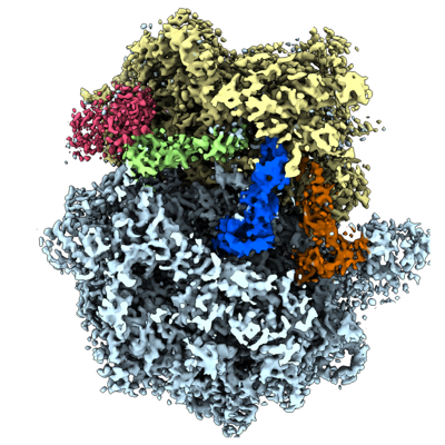
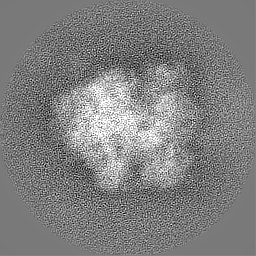
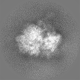
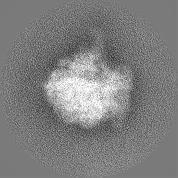
| Title | 70S ribosome with EF-Tu-tRNA, P and E-site tRNAs in chloramphenicol-treated Mycoplasma pneumoniae cells, K3 data | |||||||||
 Map data Map data | ||||||||||
 Sample Sample |
| |||||||||
 Keywords Keywords | In situ / cryo-electron tomography / bacterial ribosome / Chloramphenicol / antibiotic / RIBOSOME | |||||||||
| Biological species |  Mycoplasmoides pneumoniae M129 (bacteria) Mycoplasmoides pneumoniae M129 (bacteria) | |||||||||
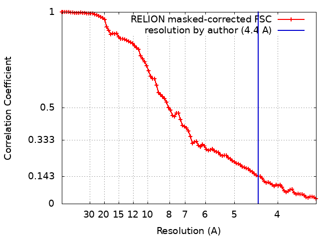
| Method | subtomogram averaging / cryo EM / Resolution: 4.4 Å | |||||||||
 Authors Authors | Xue L / Spahn C / Schacherl M / Mahamid J | |||||||||
| Funding support |  United States, United States,  Germany, 2 items Germany, 2 items
| |||||||||
 Citation Citation | Journal: Nat Struct Mol Biol / Year: 2025 Title: Structural insights into context-dependent inhibitory mechanisms of chloramphenicol in cells. Authors: Liang Xue / Christian M T Spahn / Magdalena Schacherl / Julia Mahamid /  Abstract: Ribosome-targeting antibiotics represent an important class of antimicrobial drugs. Chloramphenicol (Cm) is a well-studied ribosomal peptidyl transferase center (PTC) binder and growing evidence ...Ribosome-targeting antibiotics represent an important class of antimicrobial drugs. Chloramphenicol (Cm) is a well-studied ribosomal peptidyl transferase center (PTC) binder and growing evidence suggests that its inhibitory action depends on the sequence of the nascent peptide. How such selective inhibition on the molecular scale manifests on the cellular level remains unclear. Here, we use cryo-electron tomography to analyze the impact of Cm inside the bacterium Mycoplasma pneumoniae. By resolving the Cm-bound ribosomes to 3.0 Å, we elucidate Cm's coordination with natural nascent peptides and transfer RNAs in the PTC. We find that Cm leads to the accumulation of a number of translation elongation states, indicating ongoing futile accommodation cycles, and to extensive ribosome collisions. We, thus, suggest that, beyond its direct inhibition of protein synthesis, the action of Cm may involve the activation of cellular stress responses. This work exemplifies how in-cell structural biology can expand the understanding of mechanisms of action for extensively studied antibiotics. | |||||||||
| History |
|
- Structure visualization
| Supplemental images |
|---|
- Downloads & links
-EMDB archive
| Map data | emd_17141.map.gz | 7.8 MB |  EMDB map data format EMDB map data format | |
|---|---|---|---|---|
| Header (meta data) | emd-17141-v30.xmlemd-17141.xml | 19.6 KB 19.6 KB | Display Display | EMDB header |
| FSC (resolution estimation) | emd_17141_fsc.xml | 9.1 KB | Display | FSC data file |
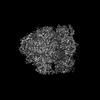
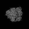
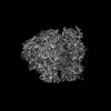
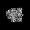
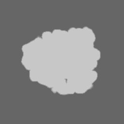
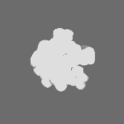
| Images |  emd_17141.png emd_17141.png | 223.7 KB | ||
| Masks | emd_17141_msk_1.map | 64 MB | Mask map | |
| Filedesc metadata | emd-17141.cif.gz | 5.4 KB | ||
| Others | emd_17141_half_map_1.map.gzemd_17141_half_map_2.map.gz | 49.7 MB 49.7 MB | ||
| Archive directory |  http://ftp.pdbj.org/pub/emdb/structures/EMD-17141ftp://ftp.pdbj.org/pub/emdb/structures/EMD-17141 http://ftp.pdbj.org/pub/emdb/structures/EMD-17141ftp://ftp.pdbj.org/pub/emdb/structures/EMD-17141 | HTTPS FTP |
-Related structure data


-Links
| EMDB pages | EMDB (EBI/PDBe) / EMDataResource |
|---|
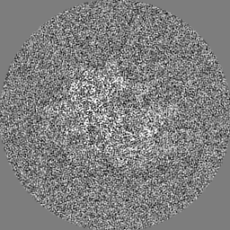
-Map
| File | Download / File: emd_17141.map.gz / Format: CCP4 / Size: 64 MB / Type: IMAGE STORED AS FLOATING POINT NUMBER (4 BYTES) | ||||||||||||||||||||||||||||||||||||
|---|---|---|---|---|---|---|---|---|---|---|---|---|---|---|---|---|---|---|---|---|---|---|---|---|---|---|---|---|---|---|---|---|---|---|---|---|---|
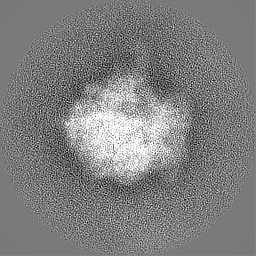
| Projections & slices | Image control
Images are generated by Spider. | ||||||||||||||||||||||||||||||||||||
| Voxel size | X=Y=Z: 1.7005 Å | ||||||||||||||||||||||||||||||||||||
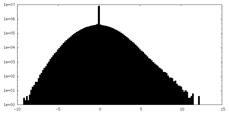
| Density |
| ||||||||||||||||||||||||||||||||||||
| Symmetry | Space group: 1 | ||||||||||||||||||||||||||||||||||||
| Details | EMDB XML:
|
 Z (Sec.)
 Y (Row.)
 X (Col.)


-Supplemental data
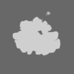
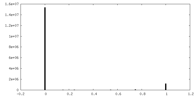
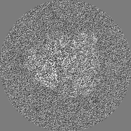
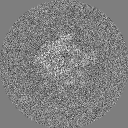
-Mask #1
| File | emd_17141_msk_1.map | ||||||||||||
|---|---|---|---|---|---|---|---|---|---|---|---|---|---|
| Projections & Slices |
| ||||||||||||
| Density Histograms |

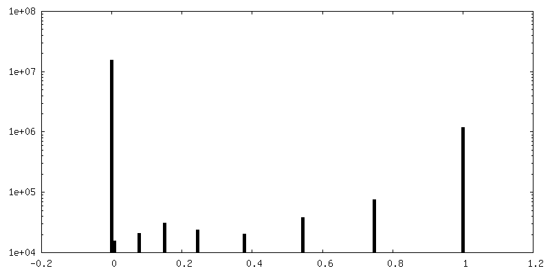
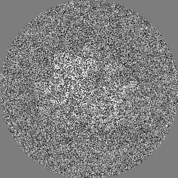
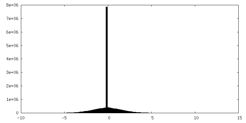
-Half map: #1
| File | emd_17141_half_map_1.map | ||||||||||||
|---|---|---|---|---|---|---|---|---|---|---|---|---|---|
| Projections & Slices |
| ||||||||||||
| Density Histograms |


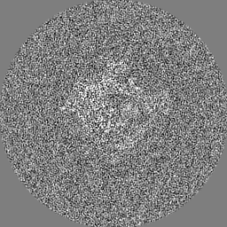

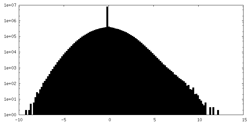
-Half map: #2
| File | emd_17141_half_map_2.map | ||||||||||||
|---|---|---|---|---|---|---|---|---|---|---|---|---|---|
| Projections & Slices |
| ||||||||||||
| Density Histograms |
- Sample components
-Entire : Mycoplasma pneumoniae M129 cells treated with chloramphenicol
| Entire | Name: Mycoplasma pneumoniae M129 cells treated with chloramphenicol |
|---|---|
| Components |
|
-Supramolecule #1: Mycoplasma pneumoniae M129 cells treated with chloramphenicol
| Supramolecule | Name: Mycoplasma pneumoniae M129 cells treated with chloramphenicol type: cell / ID: 1 / Parent: 0 |
|---|---|
| Source (natural) | Organism: Mycoplasmoides pneumoniae M129 (bacteria) |
-Experimental details
-Structure determination
| Method | cryo EM |
|---|---|
 Processing Processing | subtomogram averaging |
| Aggregation state | cell |
-Sample preparation
| Buffer | pH: 7.4 Details: Modified Hayflick medium: 14.7g/l Difco PPLO(Becton Dickinson), 20% (v/v) Gibco horse serum (New Zealand origin), 100 mM HEPES-Na; pH 7.4, 1% (w/w) glucose, 0.002% (w/w) phenol red, 1000 U/ml penicillin G. |
|---|---|
| Grid | Model: Quantifoil R2/1 / Material: GOLD / Mesh: 200 / Support film - Material: CARBON / Support film - topology: HOLEY / Pretreatment - Type: GLOW DISCHARGE |
| Vitrification | Cryogen name: ETHANE-PROPANE / Instrument: HOMEMADE PLUNGER Details: Back-side blotting for 2-3 seconds before plunging using a manual plunger without an environmental control chamber.. |
| Details | Mycoplasma pneumoniae M129 cells were grown on gold Quantifoil grids at 37 Celsius in modified Hayflick medium. Treatment with chloramphenicol at a final concentration of 0.2 mg/ml was performed for approximately 15 minutes before plunge freezing. |
- Electron microscopy
| Microscope | FEI TITAN KRIOS |
|---|---|
| Specialist optics | Energy filter - Name: GIF Bioquantum / Energy filter - Slit width: 20 eV |
| Image recording | Film or detector model: GATAN K3 BIOQUANTUM (6k x 4k) / Number real images: 1 / Average electron dose: 3.34 e/Å2 Details: Gatan K3 camera in non-CDS counting mode, targeted dose rate on camera ~20 e/pixel/second, 10 frames per tilt image, constant exposure time for each tilt, pixel size 1.329A |
| Electron beam | Acceleration voltage: 300 kV / Electron source:  FIELD EMISSION GUN FIELD EMISSION GUN |
| Electron optics | Illumination mode: FLOOD BEAM / Imaging mode: BRIGHT FIELD / Cs: 2.7 mm / Nominal defocus max: 3.25 µm / Nominal defocus min: 1.0 µm / Nominal magnification: 64000 |
| Sample stage | Specimen holder model: FEI TITAN KRIOS AUTOGRID HOLDER / Cooling holder cryogen: NITROGEN |
| Experimental equipment |  Model: Titan Krios / Image courtesy: FEI Company |