 Movie
Movie Controller


+ Open data

- Basic information
| Entry | Database: PDB / ID: 8kbz | |||||||||
|---|---|---|---|---|---|---|---|---|---|---|
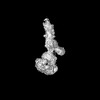
| Title | Cryo-EM structure of human ATG9A in LMNG micelles | |||||||||
 Components Components | Autophagy-related protein 9A | |||||||||
 Keywords Keywords | MEMBRANE PROTEIN / Lipid scramblase / ATG9A / single particle cryo-EM | |||||||||
| Function / homology |  Function and homology information Function and homology informationprogrammed necrotic cell death / phospholipid scramblase activity / protein localization to phagophore assembly site / phagophore assembly site membrane / piecemeal microautophagy of the nucleus / bone morphogenesis / phagophore assembly site / reticulophagy / Macroautophagy / autophagosome assembly ...programmed necrotic cell death / phospholipid scramblase activity / protein localization to phagophore assembly site / phagophore assembly site membrane / piecemeal microautophagy of the nucleus / bone morphogenesis / phagophore assembly site / reticulophagy / Macroautophagy / autophagosome assembly / mitophagy / autophagosome / synaptic membrane / PINK1-PRKN Mediated Mitophagy / trans-Golgi network / recycling endosome / mitochondrial membrane / recycling endosome membrane / late endosome / late endosome membrane / endosome / Golgi membrane / endoplasmic reticulum membrane / Golgi apparatus / mitochondrion / membrane Similarity search - Function | |||||||||
| Biological species |  Homo sapiens (human) Homo sapiens (human) | |||||||||
| Method | ELECTRON MICROSCOPY / single particle reconstruction / cryo EM / Resolution: 3.97 Å | |||||||||
 Authors Authors | Yang, W. / Goran, S. | |||||||||
| Funding support |  China, 2items China, 2items
| |||||||||
 Citation Citation | Journal: Nat Struct Mol Biol / Year: 2025 Title: Structural basis for lipid transfer by the ATG2A-ATG9A complex. Authors: Yang Wang / Selma Dahmane / Rujuan Ti / Xinyi Mai / Lizhe Zhu / Lars-Anders Carlson / Goran Stjepanovic /  Abstract: Autophagy is characterized by the formation of double-membrane vesicles called autophagosomes. Autophagy-related proteins (ATGs) 2A and 9A have an essential role in autophagy by mediating lipid ...Autophagy is characterized by the formation of double-membrane vesicles called autophagosomes. Autophagy-related proteins (ATGs) 2A and 9A have an essential role in autophagy by mediating lipid transfer and re-equilibration between membranes for autophagosome formation. Here we report the cryo-electron microscopy structures of human ATG2A in complex with WD-repeat protein interacting with phosphoinositides 4 (WIPI4) at 3.2 Å and the ATG2A-WIPI4-ATG9A complex at 7 Å global resolution. On the basis of molecular dynamics simulations, we propose a mechanism of lipid extraction from the donor membranes. Our analysis revealed 3:1 stoichiometry of the ATG9A-ATG2A complex, directly aligning the ATG9A lateral pore with ATG2A lipid transfer cavity, and an interaction of the ATG9A trimer with both the N-terminal and the C-terminal tip of rod-shaped ATG2A. Cryo-electron tomography of ATG2A liposome-binding states showed that ATG2A tethers lipid vesicles at different orientations. In summary, this study provides a molecular basis for the growth of the phagophore membrane and lends structural insights into spatially coupled lipid transport and re-equilibration during autophagosome formation. | |||||||||
| History |
|
- Structure visualization
| Structure viewer | Molecule: MolmilJmol/JSmol |
|---|
- Downloads & links
-Download
| PDBx/mmCIF format | 8kbz.cif.gz | 244.9 KB | Display | PDBx/mmCIF format |
|---|---|---|---|---|
| PDB format | pdb8kbz.ent.gz | 172.8 KB | Display | PDB format |
| PDBx/mmJSON format | 8kbz.json.gz | Tree view | PDBx/mmJSON format | |
| Others |  Other downloads Other downloads |
-Validation report
| Arichive directory | https://data.pdbj.org/pub/pdb/validation_reports/kb/8kbzftp://data.pdbj.org/pub/pdb/validation_reports/kb/8kbz | HTTPS FTP |
|---|
-Related structure data
| Related structure data | 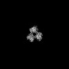 37088MC  8kbxC 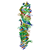 8kbyC 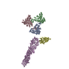 8kc3C  8y1lC M: map data used to model this data C: citing same article ( |
|---|---|
| Similar structure data |


-Links
 PDBj
PDBj

- Assembly
| Deposited unit | 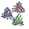
|
|---|---|
| 1 |
|
-Components
| #1: Protein | Mass: 94551.031 Da / Num. of mol.: 3 Source method: isolated from a genetically manipulated source Source: (gene. exp.) Homo sapiens (human) / Gene: ATG9A, APG9L1 / Production host: Homo sapiens (human) / References: UniProt: Q7Z3C6Has protein modification | N | |
|---|
-Experimental details
-Experiment
| Experiment | Method: ELECTRON MICROSCOPY |
|---|---|
| EM experiment | Aggregation state: PARTICLE / 3D reconstruction method: single particle reconstruction |
- Sample preparation
| Component | Name: ATG9A trimer / Type: COMPLEX / Entity ID: all / Source: MULTIPLE SOURCES |
|---|---|
| Molecular weight | Value: 0.283 MDa / Experimental value: NO |
| Source (natural) | Organism: Homo sapiens (human) |
| Source (recombinant) | Organism: Homo sapiens (human) |
| Buffer solution | pH: 8 |
| Specimen | Embedding applied: NO / Shadowing applied: NO / Staining applied: NO / Vitrification applied: YES |
| Vitrification | Cryogen name: ETHANE |
- Electron microscopy imaging
| Experimental equipment |  Model: Titan Krios / Image courtesy: FEI Company |
|---|---|
| Microscopy | Model: FEI TITAN KRIOS |
| Electron gun | Electron source:  FIELD EMISSION GUN / Accelerating voltage: 300 kV / Illumination mode: FLOOD BEAM FIELD EMISSION GUN / Accelerating voltage: 300 kV / Illumination mode: FLOOD BEAM |
| Electron lens | Mode: BRIGHT FIELD / Nominal magnification: 105000 X / Nominal defocus max: 1900 nm / Nominal defocus min: 1400 nm / C2 aperture diameter: 100 µm |
| Image recording | Electron dose: 65.17 e/Å2 / Film or detector model: GATAN K3 (6k x 4k) |
- Processing
| CTF correction | Type: PHASE FLIPPING AND AMPLITUDE CORRECTION |
|---|---|
| 3D reconstruction | Resolution: 3.97 Å / Resolution method: FSC 0.143 CUT-OFF / Num. of particles: 224219 / Symmetry type: POINT |
