Movie
Movie Controller

[English] 日本語
 Yorodumi
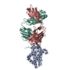
- PDB-8jn1: Cryo-EM structure of dengue virus serotype 3 strain EHIE46200Y19 ... -
+ Open data

- Basic information
| Entry | Database: PDB / ID: 8jn1 | |||||||||||||||||||||||||||||||||||||||
|---|---|---|---|---|---|---|---|---|---|---|---|---|---|---|---|---|---|---|---|---|---|---|---|---|---|---|---|---|---|---|---|---|---|---|---|---|---|---|---|---|
| Title | Cryo-EM structure of dengue virus serotype 3 strain EHIE46200Y19 in complex with human antibody DENV-115 IgG at 4 deg C (subparticle LLR-LRR) | |||||||||||||||||||||||||||||||||||||||
 Components Components |
| |||||||||||||||||||||||||||||||||||||||
 Keywords Keywords | VIRUS / dengue virus / human antibody / dengue-antibody structure | |||||||||||||||||||||||||||||||||||||||
| Function / homology |  Function and homology information Function and homology informationclathrin-dependent endocytosis of virus by host cell / protein dimerization activity / symbiont-mediated suppression of host innate immune response / host cell endoplasmic reticulum membrane / fusion of virus membrane with host endosome membrane / viral envelope / virion attachment to host cell / virion membrane / extracellular region Similarity search - Function | |||||||||||||||||||||||||||||||||||||||
| Biological species |  Dengue virus type 3 Dengue virus type 3 Homo sapiens (human) Homo sapiens (human) | |||||||||||||||||||||||||||||||||||||||
| Method | ELECTRON MICROSCOPY / single particle reconstruction / cryo EM / Resolution: 3.5 Å | |||||||||||||||||||||||||||||||||||||||
 Authors Authors | Fibriansah, G. / Ng, T.S. / Tan, A.W.K. / Shi, J. / Lok, S.M. | |||||||||||||||||||||||||||||||||||||||
| Funding support |  Singapore, 1items Singapore, 1items
| |||||||||||||||||||||||||||||||||||||||
 Citation Citation | Journal: Nat Commun / Year: 2025 Title: Ultrapotent human antibodies lock E protein dimers central region of diverse DENV3 morphological variants. Authors: Guntur Fibriansah / Thiam-Seng Ng / Xin-Ni Lim / Anastasia Shebanova / Lee Ching Ng / Song Ling Tan / Aaron W K Tan / Jian Shi / James E Crowe / Shee-Mei Lok /  Abstract: Dengue virus (DENV) consists of four serotypes (DENV1-4). Current vaccines induce differing levels of immune response against the four serotypes, that might prime recipients to develop severe disease ...Dengue virus (DENV) consists of four serotypes (DENV1-4). Current vaccines induce differing levels of immune response against the four serotypes, that might prime recipients to develop severe disease in subsequent infections. Several DENV tetravalent vaccine clinical trials suggested an increased incidence in severe DENV3 cases, suggesting a need to develop DENV3 therapeutics. Human monoclonal antibodies (HMAbs) DENV-290 and DENV-115 are ultrapotent against diverse DENV3 strains with differing particle morphologies. They mainly neutralize by inhibition of virus attachment to cells. CryoEM structures of Fabs complexed with differing DENV3 morphological variants show their Fabs binding across two E protein protomers at the center of the E dimer. This new class of E protein dimer binding antibodies is named EDE-C. The cryoEM structures also show how IgGs engage the DENV particles. Results define the structural and molecular basis for the ultrapotent activity of EDE-C antibodies. | |||||||||||||||||||||||||||||||||||||||
| History |
|
- Structure visualization
| Structure viewer | Molecule: MolmilJmol/JSmol |
|---|
- Downloads & links
-Download
| PDBx/mmCIF format | 8jn1.cif.gz | 331.8 KB | Display | PDBx/mmCIF format |
|---|---|---|---|---|
| PDB format | pdb8jn1.ent.gz | 272.9 KB | Display | PDB format |
| PDBx/mmJSON format | 8jn1.json.gz | Tree view | PDBx/mmJSON format | |
| Others |  Other downloads Other downloads |
-Validation report
| Arichive directory | https://data.pdbj.org/pub/pdb/validation_reports/jn/8jn1ftp://data.pdbj.org/pub/pdb/validation_reports/jn/8jn1 | HTTPS FTP |
|---|
-Related structure data
| Related structure data |  36429MC  8jn2C  8jn3C  8jn4C  8jn5C  8jn6C  8jn7C C: citing same article ( M: map data used to model this data |
|---|---|
| Similar structure data |


-Links
 PDBj
PDBj


- Assembly
| Deposited unit | 
|
|---|---|
| 1 | 
|
| 2 |
|
| 3 | 
|
| Symmetry | Point symmetry: (Schoenflies symbol: C2 (2 fold cyclic)) |
-Components
-Protein , 2 types, 6 molecules ACEBDF
| #1: Protein | Mass: 53731.734 Da / Num. of mol.: 3 Source method: isolated from a genetically manipulated source Source: (gene. exp.) Dengue virus type 3 / Production host:  #2: Protein | Mass: 8365.854 Da / Num. of mol.: 3 Source method: isolated from a genetically manipulated source Source: (gene. exp.) Dengue virus type 3 / Gene: PrM/M, E / Production host: |
|---|
-Antibody , 2 types, 2 molecules HL
| #3: Antibody | Mass: 13607.289 Da / Num. of mol.: 1 Source method: isolated from a genetically manipulated source Source: (gene. exp.) Homo sapiens (human) / Cell (production host): EXPI293F / Cell line (production host): EXPI293F / Production host: Homo sapiens (human) |
|---|---|
| #4: Antibody | Mass: 11496.620 Da / Num. of mol.: 1 Source method: isolated from a genetically manipulated source Source: (gene. exp.) Homo sapiens (human) / Cell (production host): EXPI293F / Cell line (production host): EXPI293F / Production host: Homo sapiens (human) |
-Sugars , 2 types, 6 molecules
| #5: Polysaccharide | 2-acetamido-2-deoxy-beta-D-glucopyranose-(1-4)-2-acetamido-2-deoxy-beta-D-glucopyranose Source method: isolated from a genetically manipulated source #6: Polysaccharide | Source method: isolated from a genetically manipulated source |
|---|
-Details
| Has ligand of interest | N |
|---|---|
| Has protein modification | Y |
-Experimental details
-Experiment
| Experiment | Method: ELECTRON MICROSCOPY |
|---|---|
| EM experiment | Aggregation state: PARTICLE / 3D reconstruction method: single particle reconstruction |
- Sample preparation
| Component |
| ||||||||||||||||||||||||
|---|---|---|---|---|---|---|---|---|---|---|---|---|---|---|---|---|---|---|---|---|---|---|---|---|---|
| Molecular weight |
| ||||||||||||||||||||||||
| Source (natural) |
| ||||||||||||||||||||||||
| Details of virus |
| ||||||||||||||||||||||||
| Buffer solution | pH: 8 Details: NTE buffer (12 mM Tris-HCl pH 8.0, 120 mM NaCl and 1 mM EDTA) | ||||||||||||||||||||||||
| Specimen | Embedding applied: NO / Shadowing applied: NO / Staining applied: NO / Vitrification applied: YES Details: The purified virus was mixed with DENV-115 IgG at a molar ratio of 1.5 Fab for every E-protein and then the mixture was incubated at 4 deg C for 30 min. | ||||||||||||||||||||||||
| Vitrification | Cryogen name: ETHANE |
- Electron microscopy imaging
| Experimental equipment |  Model: Titan Krios / Image courtesy: FEI Company |
|---|---|
| Microscopy | Model: FEI TITAN KRIOS |
| Electron gun | Electron source:  FIELD EMISSION GUN / Accelerating voltage: 300 kV / Illumination mode: FLOOD BEAM FIELD EMISSION GUN / Accelerating voltage: 300 kV / Illumination mode: FLOOD BEAM |
| Electron lens | Mode: BRIGHT FIELD / Nominal defocus max: 4600 nm / Nominal defocus min: 400 nm |
| Image recording | Electron dose: 22.8 e/Å2 / Film or detector model: GATAN K3 (6k x 4k) |
- Processing
| EM software |
| ||||||||||||||||||||||||||||
|---|---|---|---|---|---|---|---|---|---|---|---|---|---|---|---|---|---|---|---|---|---|---|---|---|---|---|---|---|---|
| CTF correction | Type: PHASE FLIPPING AND AMPLITUDE CORRECTION | ||||||||||||||||||||||||||||
| Symmetry | Point symmetry: C2 (2 fold cyclic) | ||||||||||||||||||||||||||||
| 3D reconstruction | Resolution: 3.5 Å / Resolution method: FSC 0.143 CUT-OFF / Num. of particles: 181866 / Symmetry type: POINT | ||||||||||||||||||||||||||||
| Atomic model building | Protocol: FLEXIBLE FIT / Space: REAL | ||||||||||||||||||||||||||||
| Atomic model building |
| ||||||||||||||||||||||||||||
| Refine LS restraints |
|