Movie
Movie Controller


+ Open data

- Basic information
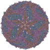
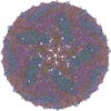
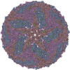
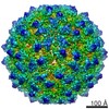
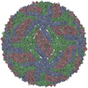
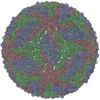
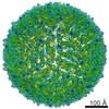
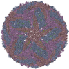
| Entry | Database: PDB / ID: 3j6s | ||||||
|---|---|---|---|---|---|---|---|
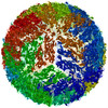
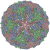
| Title | Cryo-EM structure of Dengue virus serotype 3 at 28 degrees C | ||||||
 Components Components |
| ||||||
 Keywords Keywords | VIRUS / Dengue virus | ||||||
| Function / homology |  Function and homology information Function and homology informationflavivirin / host cell mitochondrion / symbiont-mediated suppression of host JAK-STAT cascade via inhibition of host TYK2 activity / symbiont-mediated suppression of host JAK-STAT cascade via inhibition of STAT2 activity / symbiont-mediated suppression of host cytoplasmic pattern recognition receptor signaling pathway via inhibition of MAVS activity / viral capsid / ribonucleoside triphosphate phosphatase activity / nucleoside-triphosphate phosphatase / double-stranded RNA binding / channel activity ...flavivirin / host cell mitochondrion / symbiont-mediated suppression of host JAK-STAT cascade via inhibition of host TYK2 activity / symbiont-mediated suppression of host JAK-STAT cascade via inhibition of STAT2 activity / symbiont-mediated suppression of host cytoplasmic pattern recognition receptor signaling pathway via inhibition of MAVS activity / viral capsid / ribonucleoside triphosphate phosphatase activity / nucleoside-triphosphate phosphatase / double-stranded RNA binding / channel activity / monoatomic ion transmembrane transport / clathrin-dependent endocytosis of virus by host cell / mRNA (guanine-N7)-methyltransferase / methyltransferase cap1 / methyltransferase cap1 activity / mRNA 5'-cap (guanine-N7-)-methyltransferase activity / RNA helicase activity / protein dimerization activity / host cell perinuclear region of cytoplasm / host cell endoplasmic reticulum membrane / RNA helicase / symbiont-mediated suppression of host type I interferon-mediated signaling pathway / serine-type endopeptidase activity / symbiont-mediated activation of host autophagy / RNA-directed RNA polymerase / viral RNA genome replication / RNA-directed RNA polymerase activity / fusion of virus membrane with host endosome membrane / viral envelope / lipid binding / virion attachment to host cell / host cell nucleus / virion membrane / structural molecule activity / ATP hydrolysis activity / proteolysis / extracellular region / ATP binding / metal ion binding Similarity search - Function | ||||||
| Biological species |  Dengue virus 3 Dengue virus 3 | ||||||
| Method | ELECTRON MICROSCOPY / single particle reconstruction / cryo EM / Resolution: 6 Å | ||||||
 Authors Authors | Fibriansah, G. / Tan, J.L. / Smith, S.A. / de Alwis, R. / Ng, T.-S. / Kostyuchenko, V.A. / Kukkaro, P. / de Silva, A.M. / Crowe Jr., J.E. / Lok, S.-M. | ||||||
 Citation Citation | Journal: Nat Commun / Year: 2015 Title: A highly potent human antibody neutralizes dengue virus serotype 3 by binding across three surface proteins. Authors: Guntur Fibriansah / Joanne L Tan / Scott A Smith / Ruklanthi de Alwis / Thiam-Seng Ng / Victor A Kostyuchenko / Ramesh S Jadi / Petra Kukkaro / Aravinda M de Silva / James E Crowe / Shee-Mei Lok /   Abstract: Dengue virus (DENV) infects ~400 million people annually. There is no licensed vaccine or therapeutic drug. Only a small fraction of the total DENV-specific antibodies in a naturally occurring dengue ...Dengue virus (DENV) infects ~400 million people annually. There is no licensed vaccine or therapeutic drug. Only a small fraction of the total DENV-specific antibodies in a naturally occurring dengue infection consists of highly neutralizing antibodies. Here we show that the DENV-specific human monoclonal antibody 5J7 is exceptionally potent, neutralizing 50% of virus at nanogram-range antibody concentration. The 9 Å resolution cryo-electron microscopy structure of the Fab 5J7-DENV complex shows that a single Fab molecule binds across three envelope proteins and engages three functionally important domains, each from a different envelope protein. These domains are critical for receptor binding and fusion to the endosomal membrane. The ability to bind to multiple domains allows the antibody to fully coat the virus surface with only 60 copies of Fab, that is, half the amount compared with other potent antibodies. Our study reveals a highly efficient and unusual mechanism of molecular recognition by an antibody. | ||||||
| History |
|
- Structure visualization
| Movie |
Movie viewer |
|---|---|
| Structure viewer | Molecule: MolmilJmol/JSmol |
- Downloads & links
-Download
| PDBx/mmCIF format | 3j6s.cif.gz | 61.4 KB | Display | PDBx/mmCIF format |
|---|---|---|---|---|
| PDB format | pdb3j6s.ent.gz | 35.7 KB | Display | PDB format |
| PDBx/mmJSON format | 3j6s.json.gz | Tree view | PDBx/mmJSON format | |
| Others |  Other downloads Other downloads |
-Validation report
| Arichive directory | https://data.pdbj.org/pub/pdb/validation_reports/j6/3j6sftp://data.pdbj.org/pub/pdb/validation_reports/j6/3j6s | HTTPS FTP |
|---|
-Related structure data
| Related structure data | 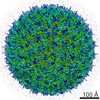 5933MC 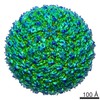 5934C  5935C  3j6tC  3j6uC M: map data used to model this data C: citing same article ( |
|---|---|
| Similar structure data |

-Links
 PDBj
PDBj


- Assembly
| Deposited unit | 
|
|---|---|
| 1 | x 60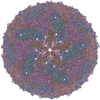
|
| 2 |
|
| 3 | x 5
|
| 4 | x 6
|
| 5 | 
|
| Symmetry | Point symmetry: (Schoenflies symbol: I (icosahedral)) |
-Components
| #1: Protein | Mass: 53682.484 Da / Num. of mol.: 3 / Fragment: UNP residues 281-773 Source method: isolated from a genetically manipulated source Source: (gene. exp.) Dengue virus 3 / Strain: D3/SG/05K863DK1/2005 / Cell line (production host): C6/36 / Production host:  #2: Protein | Mass: 8347.836 Da / Num. of mol.: 3 / Fragment: UNP residues 206-280 Source method: isolated from a genetically manipulated source Source: (gene. exp.) Dengue virus 3 / Strain: D3/SG/05K863DK1/2005 / Cell line (production host): C6/36 / Production host: |
|---|
-Experimental details
-Experiment
| Experiment | Method: ELECTRON MICROSCOPY |
|---|---|
| EM experiment | Aggregation state: PARTICLE / 3D reconstruction method: single particle reconstruction |
- Sample preparation
| Component | Name: Dengue virus serotype 3 / Type: VIRUS |
|---|---|
| Details of virus | Empty: NO / Enveloped: YES / Host category: VERTEBRATES / Isolate: STRAIN / Type: VIRION |
| Natural host | Organism: Homo sapiens |
| Buffer solution | Name: 10 mM Tris-HCl, pH 8.0, 120 mM NaCl, 1 mM EDTA / pH: 8 / Details: 10 mM Tris-HCl, pH 8.0, 120 mM NaCl, 1 mM EDTA |
| Specimen | Embedding applied: NO / Shadowing applied: NO / Staining applied: NO / Vitrification applied: YES |
| Specimen support | Details: ultra-thin carbon-coated lacey carbon grid |
| Vitrification | Instrument: FEI VITROBOT MARK IV / Cryogen name: ETHANE / Temp: 100 K / Humidity: 100 % Details: Blotted with filter paper for 2 seconds prior to snap freezing in liquid ethane (FEI VITROBOT MARK IV) Method: Blotted with filter paper for 2 seconds prior to snap freezing |
- Electron microscopy imaging
| Experimental equipment |  Model: Titan Krios / Image courtesy: FEI Company |
|---|---|
| Microscopy | Model: FEI TITAN KRIOS / Date: Oct 27, 2011 |
| Electron gun | Electron source:  FIELD EMISSION GUN / Accelerating voltage: 300 kV / Illumination mode: SPOT SCAN FIELD EMISSION GUN / Accelerating voltage: 300 kV / Illumination mode: SPOT SCAN |
| Electron lens | Mode: BRIGHT FIELD / Nominal magnification: 75000 X / Nominal defocus max: 3500 nm / Nominal defocus min: 1100 nm / Cs: 2.7 mm / Camera length: 0 mm |
| Specimen holder | Specimen holder model: FEI TITAN KRIOS AUTOGRID HOLDER / Temperature: 100 K / Tilt angle max: 0 ° / Tilt angle min: 0 ° |
| Image recording | Electron dose: 20.4 e/Å2 / Film or detector model: GATAN ULTRASCAN 4000 (4k x 4k) |
| Image scans | Num. digital images: 836 |
| Radiation | Protocol: SINGLE WAVELENGTH / Monochromatic (M) / Laue (L): M / Scattering type: x-ray |
| Radiation wavelength | Relative weight: 1 |
- Processing
| EM software |
| ||||||||||||||||||||||||||||||||
|---|---|---|---|---|---|---|---|---|---|---|---|---|---|---|---|---|---|---|---|---|---|---|---|---|---|---|---|---|---|---|---|---|---|
| CTF correction | Details: each particle | ||||||||||||||||||||||||||||||||
| Symmetry | Point symmetry: I (icosahedral) | ||||||||||||||||||||||||||||||||
| 3D reconstruction | Method: Cross-common lines / Resolution: 6 Å / Resolution method: FSC 0.5 CUT-OFF / Num. of particles: 6800 / Nominal pixel size: 1.16 Å / Actual pixel size: 1.16 Å Details: (Single particle details: Particles were manually selected.) (Single particle--Applied symmetry: I) Symmetry type: POINT | ||||||||||||||||||||||||||||||||
| Atomic model building | Protocol: FLEXIBLE FIT / Space: REAL / Target criteria: real space correlation Details: REFINEMENT PROTOCOL--flexible DETAILS--Initially fitted in Chimera, model rebuilt in Coot, refined in NAMD/MDFF | ||||||||||||||||||||||||||||||||
| Atomic model building | 3D fitting-ID: 1 / Accession code: 3J27 / Initial refinement model-ID: 1 / PDB-ID: 3J27 / Source name: PDB / Type: experimental model
| ||||||||||||||||||||||||||||||||
| Refinement step | Cycle: LAST
|