| 登録情報 | データベース: PDB / ID: 8g24
|
|---|
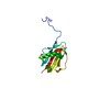
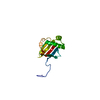
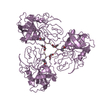
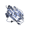
| タイトル | Crystal Structure of Cathepsin-G and Neutrophil Elastase Inhibited by S. aureus EapH2 at pH 5.5 |
|---|
 要素 要素 | - Cathepsin-G
- MAP domain-containing protein
- Neutrophil elastase
|
|---|
 キーワード キーワード | HYDROLASE/INHIBITOR / Protease Inhibitor / Immune Evasion / Neutrophil / S. aureus / HYDROLASE-INHIBITOR complex |
|---|
| 機能・相同性 |  機能・相同性情報 機能・相同性情報
cathepsin G / biofilm matrix disassembly / neutrophil-mediated killing of gram-positive bacterium / leukocyte elastase / biosynthetic process of antibacterial peptides active against Gram-negative bacteria / Expression of NOTCH2NL genes / acute inflammatory response to antigenic stimulus / neutrophil-mediated killing of fungus / negative regulation of chemotaxis / positive regulation of leukocyte tethering or rolling ...cathepsin G / biofilm matrix disassembly / neutrophil-mediated killing of gram-positive bacterium / leukocyte elastase / biosynthetic process of antibacterial peptides active against Gram-negative bacteria / Expression of NOTCH2NL genes / acute inflammatory response to antigenic stimulus / neutrophil-mediated killing of fungus / negative regulation of chemotaxis / positive regulation of leukocyte tethering or rolling / purinergic nucleotide receptor signaling pathway / caspase binding / response to yeast / negative regulation of T cell activation / leukocyte migration involved in inflammatory response / negative regulation of interleukin-8 production / negative regulation of chemokine production / neutrophil activation / Suppression of apoptosis / Interleukin-1 processing / positive regulation of platelet aggregation / Antimicrobial peptides / pyroptotic inflammatory response / cytokine binding / neutrophil-mediated killing of gram-negative bacterium / Activation of Matrix Metalloproteinases / positive regulation of MAP kinase activity / monocyte chemotaxis / Collagen degradation / extracellular matrix disassembly / Pyroptosis / phagocytosis / defense response to fungus / Purinergic signaling in leishmaniasis infection / response to UV / Metabolism of Angiotensinogen to Angiotensins / angiotensin maturation / phagocytic vesicle / Degradation of the extracellular matrix / transcription repressor complex / serine-type peptidase activity / positive regulation of smooth muscle cell proliferation / secretory granule / protein maturation / Regulation of Complement cascade / positive regulation of interleukin-8 production / protein catabolic process / positive regulation of immune response / protein processing / platelet activation / negative regulation of inflammatory response / specific granule lumen / intracellular calcium ion homeostasis / Regulation of Insulin-like Growth Factor (IGF) transport and uptake by Insulin-like Growth Factor Binding Proteins (IGFBPs) / cytokine-mediated signaling pathway / cytoplasmic stress granule / azurophil granule lumen / antibacterial humoral response / transcription corepressor activity / peptidase activity / heparin binding / : / cellular response to lipopolysaccharide / protease binding / response to lipopolysaccharide / endopeptidase activity / defense response to Gram-negative bacterium / lysosome / protein phosphorylation / defense response to Gram-positive bacterium / defense response to bacterium / immune response / receptor ligand activity / serine-type endopeptidase activity / intracellular membrane-bounded organelle / Neutrophil degranulation / cell surface / negative regulation of transcription by RNA polymerase II / Golgi apparatus / proteolysis / extracellular space / extracellular exosome / extracellular region / nucleus / membrane / plasma membrane / cytosol / cytoplasm類似検索 - 分子機能 MAP domain / MAP domain / MAP repeat profile. / : / Serine proteases, trypsin family, histidine active site / Serine proteases, trypsin family, serine active site / Serine proteases, trypsin family, histidine active site. / Peptidase S1A, chymotrypsin family / Serine proteases, trypsin family, serine active site. / Serine proteases, trypsin domain profile. ...MAP domain / MAP domain / MAP repeat profile. / : / Serine proteases, trypsin family, histidine active site / Serine proteases, trypsin family, serine active site / Serine proteases, trypsin family, histidine active site. / Peptidase S1A, chymotrypsin family / Serine proteases, trypsin family, serine active site. / Serine proteases, trypsin domain profile. / Trypsin-like serine protease / Serine proteases, trypsin domain / Trypsin / Peptidase S1, PA clan, chymotrypsin-like fold / Peptidase S1, PA clan類似検索 - ドメイン・相同性 MAP domain-containing protein / Neutrophil elastase / Cathepsin G類似検索 - 構成要素 |
|---|
| 生物種 |  Staphylococcus aureus subsp. aureus Mu50 (黄色ブドウ球菌) Staphylococcus aureus subsp. aureus Mu50 (黄色ブドウ球菌)
 Homo sapiens (ヒト) Homo sapiens (ヒト) |
|---|
| 手法 |  X線回折 / シンクロトロン / 分子置換 / 解像度: 1.82 Å X線回折 / シンクロトロン / 分子置換 / 解像度: 1.82 Å |
|---|
 データ登録者 データ登録者 | Mishra, N.B. / Geisbrecht, B.V. |
|---|
| 資金援助 |  米国, 1件 米国, 1件 | 組織 | 認可番号 | 国 |
|---|
| National Institutes of Health/National Institute of General Medical Sciences (NIH/NIGMS) | GM140852 | 米国 |
|
|---|
 引用 引用 | ジャーナル: J.Biol.Chem. / 年: 2023
タイトル: Simultaneous inhibition of two neutrophil serine proteases by the S. aureus innate immune evasion protein EapH2.
著者: Mishra, N. / Herdendorf, T.J. / Prakash, O. / Geisbrecht, B.V. |
|---|
| 履歴 | | 登録 | 2023年2月3日 | 登録サイト: RCSB / 処理サイト: RCSB |
|---|
| 改定 1.0 | 2023年4月26日 | Provider: repository / タイプ: Initial release |
|---|
| 改定 1.1 | 2023年6月14日 | Group: Database references / カテゴリ: citation / citation_author
Item: _citation.country / _citation.journal_abbrev ..._citation.country / _citation.journal_abbrev / _citation.journal_id_ASTM / _citation.journal_id_CSD / _citation.journal_id_ISSN / _citation.page_first / _citation.page_last / _citation.pdbx_database_id_DOI / _citation.pdbx_database_id_PubMed / _citation.title / _citation.year |
|---|
| 改定 1.2 | 2023年7月19日 | Group: Database references / カテゴリ: citation / Item: _citation.journal_volume |
|---|
| 改定 1.3 | 2023年10月25日 | Group: Data collection / Refinement description
カテゴリ: chem_comp_atom / chem_comp_bond / pdbx_initial_refinement_model
Item: _pdbx_initial_refinement_model.details |
|---|
| 改定 1.4 | 2024年10月23日 | Group: Structure summary
カテゴリ: pdbx_entry_details / pdbx_modification_feature
Item: _pdbx_entry_details.has_protein_modification |
|---|
|
|---|
 ムービー
ムービー コントローラー


 データを開く
 基本情報
基本情報 構造の表示
構造の表示 ダウンロードとリンク
 その他のダウンロード


 PDBj
PDBj


 集合体
集合体
 分子量: 18.015 Da / 分子数: 163 / 由来タイプ: 天然 / 式: H2O
 試料調製
試料調製 解析
解析