 Movie
Movie Controller

[English] 日本語
 Yorodumi
- PDB-8g25: Crystal Structure of Cathepsin-G and Neutrophil Elastase Inhibite... -
+ Open data

- Basic information
| Entry | Database: PDB / ID: 8g25 | ||||||
|---|---|---|---|---|---|---|---|
| Title | Crystal Structure of Cathepsin-G and Neutrophil Elastase Inhibited by S. aureus EapH2 at pH 7.5 | ||||||
 Components Components |
| ||||||
 Keywords Keywords | HYDROLASE/INHIBITOR / Protease Inhibitor / Immune Evasion / Neutrophil / S. aureus / HYDROLASE-INHIBITOR complex | ||||||
| Function / homology |  Function and homology information Function and homology informationcathepsin G / biofilm matrix disassembly / neutrophil-mediated killing of gram-positive bacterium / leukocyte elastase / biosynthetic process of antibacterial peptides active against Gram-negative bacteria / Expression of NOTCH2NL genes / acute inflammatory response to antigenic stimulus / neutrophil-mediated killing of fungus / negative regulation of chemotaxis / positive regulation of leukocyte tethering or rolling ...cathepsin G / biofilm matrix disassembly / neutrophil-mediated killing of gram-positive bacterium / leukocyte elastase / biosynthetic process of antibacterial peptides active against Gram-negative bacteria / Expression of NOTCH2NL genes / acute inflammatory response to antigenic stimulus / neutrophil-mediated killing of fungus / negative regulation of chemotaxis / positive regulation of leukocyte tethering or rolling / purinergic nucleotide receptor signaling pathway / caspase binding / leukocyte migration involved in inflammatory response / response to yeast / negative regulation of chemokine production / neutrophil activation / negative regulation of interleukin-8 production / Suppression of apoptosis / Interleukin-1 processing / positive regulation of platelet aggregation / Antimicrobial peptides / negative regulation of T cell activation / neutrophil-mediated killing of gram-negative bacterium / Activation of Matrix Metalloproteinases / positive regulation of MAP kinase activity / cytokine binding / Collagen degradation / extracellular matrix disassembly / monocyte chemotaxis / pyroptotic inflammatory response / Pyroptosis / defense response to fungus / response to UV / Metabolism of Angiotensinogen to Angiotensins / phagocytosis / Purinergic signaling in leishmaniasis infection / Degradation of the extracellular matrix / angiotensin maturation / phagocytic vesicle / transcription repressor complex / secretory granule / positive regulation of smooth muscle cell proliferation / Regulation of Complement cascade / serine-type peptidase activity / positive regulation of interleukin-8 production / protein catabolic process / protein maturation / protein processing / platelet activation / negative regulation of inflammatory response / specific granule lumen / positive regulation of immune response / Regulation of Insulin-like Growth Factor (IGF) transport and uptake by Insulin-like Growth Factor Binding Proteins (IGFBPs) / cytokine-mediated signaling pathway / intracellular calcium ion homeostasis / cytoplasmic stress granule / azurophil granule lumen / transcription corepressor activity / peptidase activity / heparin binding / antibacterial humoral response / extracellular matrix / cellular response to lipopolysaccharide / protease binding / endopeptidase activity / response to lipopolysaccharide / defense response to Gram-negative bacterium / lysosome / defense response to bacterium / defense response to Gram-positive bacterium / immune response / receptor ligand activity / serine-type endopeptidase activity / Neutrophil degranulation / cell surface / negative regulation of transcription by RNA polymerase II / Golgi apparatus / proteolysis / : / extracellular exosome / extracellular region / membrane / nucleus / plasma membrane / cytoplasm / cytosol Similarity search - Function | ||||||
| Biological species |  Staphylococcus aureus subsp. aureus Mu50 (bacteria) Staphylococcus aureus subsp. aureus Mu50 (bacteria) Homo sapiens (human) Homo sapiens (human) | ||||||
| Method |  X-RAY DIFFRACTION / SYNCHROTRON / MOLECULAR REPLACEMENT / Resolution: 1.8 Å X-RAY DIFFRACTION / SYNCHROTRON / MOLECULAR REPLACEMENT / Resolution: 1.8 Å | ||||||
 Authors Authors | Mishra, N.B. / Geisbrecht, B.V. | ||||||
| Funding support |  United States, 1items United States, 1items
| ||||||
 Citation Citation | Journal: J.Biol.Chem. / Year: 2023 Title: Simultaneous inhibition of two neutrophil serine proteases by the S. aureus innate immune evasion protein EapH2. Authors: Mishra, N. / Herdendorf, T.J. / Prakash, O. / Geisbrecht, B.V. | ||||||
| History |
|
- Structure visualization
| Structure viewer | Molecule: MolmilJmol/JSmol |
|---|
- Downloads & links
-Download
| PDBx/mmCIF format | 8g25.cif.gz | 657.8 KB | Display | PDBx/mmCIF format |
|---|---|---|---|---|
| PDB format | pdb8g25.ent.gz | 541.8 KB | Display | PDB format |
| PDBx/mmJSON format | 8g25.json.gz | Tree view | PDBx/mmJSON format | |
| Others |  Other downloads Other downloads |
-Validation report
| Arichive directory | https://data.pdbj.org/pub/pdb/validation_reports/g2/8g25ftp://data.pdbj.org/pub/pdb/validation_reports/g2/8g25 | HTTPS FTP |
|---|
-Related structure data
| Related structure data |  8g24C  8g26C 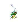 8gdgC  8gdhC  1au8S 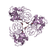 1hneS 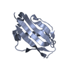 1yn5S S: Starting model for refinement C: citing same article ( |
|---|---|
| Similar structure data |
-Links
 PDBj
PDBj


- Assembly
| Deposited unit | 
| ||||||||
|---|---|---|---|---|---|---|---|---|---|
| 1 | 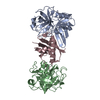
| ||||||||
| 2 | 
| ||||||||
| 3 | 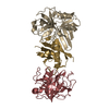
| ||||||||
| Unit cell |
|
-Components
| #1: Protein | Mass: 25397.131 Da / Num. of mol.: 3 / Fragment: C-terminal truncation (UNP residues 21-243) / Source method: isolated from a natural source / Source: (natural) Homo sapiens (human) / Tissue: neutrophil / References: UniProt: P08311#2: Protein | Mass: 23318.982 Da / Num. of mol.: 3 / Source method: isolated from a natural source / Source: (natural) Homo sapiens (human) / Tissue: neutrophil / References: UniProt: P08246, leukocyte elastase#3: Protein | Mass: 13123.912 Da / Num. of mol.: 3 Source method: isolated from a genetically manipulated source Source: (gene. exp.) Staphylococcus aureus subsp. aureus Mu50 (bacteria)Gene: SAV0981 / Production host: #4: Water | ChemComp-HOH / |  Mass: 18.015 Da / Num. of mol.: 633 / Source method: isolated from a natural source / Formula: H2O Mass: 18.015 Da / Num. of mol.: 633 / Source method: isolated from a natural source / Formula: H2OHas protein modification | Y | |
|---|
-Experimental details
-Experiment
| Experiment | Method: X-RAY DIFFRACTION / Number of used crystals: 1 |
|---|
- Sample preparation
| Crystal | Density Matthews: 2.2 Å3/Da / Density % sol: 44.06 % |
|---|---|
| Crystal grow | Temperature: 293 K / Method: vapor diffusion, hanging drop / pH: 7.5 Details: 0.1 M HEPES, 0.2 M lithium sulfate, 26% w/v PEG3350 |
-Data collection
| Diffraction | Mean temperature: 100 K / Serial crystal experiment: N | |||||||||||||||||||||||||||||||||||||||||||||||||||||||||||||||||||||||||||||
|---|---|---|---|---|---|---|---|---|---|---|---|---|---|---|---|---|---|---|---|---|---|---|---|---|---|---|---|---|---|---|---|---|---|---|---|---|---|---|---|---|---|---|---|---|---|---|---|---|---|---|---|---|---|---|---|---|---|---|---|---|---|---|---|---|---|---|---|---|---|---|---|---|---|---|---|---|---|---|
| Diffraction source | Source: SYNCHROTRON / Site: APS / Beamline: 22-ID / Wavelength: 1 Å | |||||||||||||||||||||||||||||||||||||||||||||||||||||||||||||||||||||||||||||
| Detector | Type: DECTRIS EIGER X 16M / Detector: PIXEL / Date: Dec 9, 2022 | |||||||||||||||||||||||||||||||||||||||||||||||||||||||||||||||||||||||||||||
| Radiation | Protocol: SINGLE WAVELENGTH / Monochromatic (M) / Laue (L): M / Scattering type: x-ray | |||||||||||||||||||||||||||||||||||||||||||||||||||||||||||||||||||||||||||||
| Radiation wavelength | Wavelength: 1 Å / Relative weight: 1 | |||||||||||||||||||||||||||||||||||||||||||||||||||||||||||||||||||||||||||||
| Reflection | Resolution: 1.8→50 Å / Num. obs: 149744 / % possible obs: 98.1 % / Redundancy: 3.3 % / CC1/2: 0.979 / Rpim(I) all: 0.075 / Χ2: 0.111 / Net I/σ(I): 5.1 / Num. measured all: 951665 | |||||||||||||||||||||||||||||||||||||||||||||||||||||||||||||||||||||||||||||
| Reflection shell | Diffraction-ID: 1
|
- Processing
| Software |
| ||||||||||||||||||||||||||||||||||||||||||||||||||||||||||||||||||||||||||||||||||||||||||||||||||
|---|---|---|---|---|---|---|---|---|---|---|---|---|---|---|---|---|---|---|---|---|---|---|---|---|---|---|---|---|---|---|---|---|---|---|---|---|---|---|---|---|---|---|---|---|---|---|---|---|---|---|---|---|---|---|---|---|---|---|---|---|---|---|---|---|---|---|---|---|---|---|---|---|---|---|---|---|---|---|---|---|---|---|---|---|---|---|---|---|---|---|---|---|---|---|---|---|---|---|---|
| Refinement | Method to determine structure: MOLECULAR REPLACEMENT Starting model: PDB entries 1AU8, 1HNE, & 1YN5 Resolution: 1.8→46.13 Å / SU ML: 0.23 / Cross valid method: FREE R-VALUE / σ(F): 0.17 / Phase error: 35.16 / Stereochemistry target values: ML
| ||||||||||||||||||||||||||||||||||||||||||||||||||||||||||||||||||||||||||||||||||||||||||||||||||
| Solvent computation | Shrinkage radii: 0.9 Å / VDW probe radii: 1.11 Å / Solvent model: FLAT BULK SOLVENT MODEL | ||||||||||||||||||||||||||||||||||||||||||||||||||||||||||||||||||||||||||||||||||||||||||||||||||
| Refinement step | Cycle: LAST / Resolution: 1.8→46.13 Å
| ||||||||||||||||||||||||||||||||||||||||||||||||||||||||||||||||||||||||||||||||||||||||||||||||||
| Refine LS restraints |
| ||||||||||||||||||||||||||||||||||||||||||||||||||||||||||||||||||||||||||||||||||||||||||||||||||
| LS refinement shell |
|
