 Movie
Movie Controller

[English] 日本語
 Yorodumi
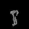
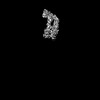
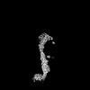
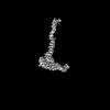
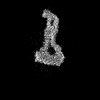
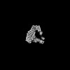
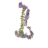
- PDB-8ftu: Crystal structure of the SNARE Use1 bound to Dsl1 complex subunit... -
+ Open data

- Basic information
| Entry | Database: PDB / ID: 8ftu | ||||||||||||
|---|---|---|---|---|---|---|---|---|---|---|---|---|---|
| Title | Crystal structure of the SNARE Use1 bound to Dsl1 complex subunits Sec39 and Dsl1, Revised Use1 structure | ||||||||||||
 Components Components |
| ||||||||||||
 Keywords Keywords | TRANSPORT PROTEIN / membrane trafficking / SNARE protein / COPI / vesicle / multisubunit tethering complex / Dsl1 complex / CATCHR complex | ||||||||||||
| Function / homology |  Function and homology information Function and homology informationRZZ complex / SNARE complex / SNAP receptor activity / retrograde vesicle-mediated transport, Golgi to endoplasmic reticulum / mitotic spindle assembly checkpoint signaling / endoplasmic reticulum to Golgi vesicle-mediated transport / protein transport / endoplasmic reticulum / cytoplasm Similarity search - Function | ||||||||||||
| Biological species |  Kluyveromyces lactis NRRL Y-1140 (yeast) Kluyveromyces lactis NRRL Y-1140 (yeast) | ||||||||||||
| Method |  X-RAY DIFFRACTION / SYNCHROTRON / MOLECULAR REPLACEMENT / Resolution: 5.73 Å X-RAY DIFFRACTION / SYNCHROTRON / MOLECULAR REPLACEMENT / Resolution: 5.73 Å | ||||||||||||
 Authors Authors | Travis, S.M. / Jeffrey, P.D. / Hughson, F.M. | ||||||||||||
| Funding support |  United States, 3items United States, 3items
| ||||||||||||
 Citation Citation | Journal: Nat Struct Mol Biol / Year: 2024 Title: Structure of a membrane tethering complex incorporating multiple SNAREs. Authors: Kevin A DAmico / Abigail E Stanton / Jaden D Shirkey / Sophie M Travis / Philip D Jeffrey / Frederick M Hughson / Abstract: Most membrane fusion reactions in eukaryotic cells are mediated by multisubunit tethering complexes (MTCs) and SNARE proteins. MTCs are much larger than SNAREs and are thought to mediate the initial ...Most membrane fusion reactions in eukaryotic cells are mediated by multisubunit tethering complexes (MTCs) and SNARE proteins. MTCs are much larger than SNAREs and are thought to mediate the initial attachment of two membranes. Complementary SNAREs then form membrane-bridging complexes whose assembly draws the membranes together for fusion. Here we present a cryo-electron microscopy structure of the simplest known MTC, the 255-kDa Dsl1 complex of Saccharomyces cerevisiae, bound to the two SNAREs that anchor it to the endoplasmic reticulum. N-terminal domains of the SNAREs form an integral part of the structure, stabilizing a Dsl1 complex configuration with unexpected similarities to the 850-kDa exocyst MTC. The structure of the SNARE-anchored Dsl1 complex and its comparison with exocyst reveal what are likely to be common principles underlying MTC function. Our structure also implies that tethers and SNAREs can work together as a single integrated machine. #1: Journal: J Biol Chem / Year: 2020Title: Structural basis for the binding of SNAREs to the multisubunit tethering complex Dsl1. Authors: Travis, S.M. / DAmico, K. / Yu, I.M. / McMahon, C. / Hamid, S. / Ramirez-Arellano, G. / Jeffrey, P.D. / Hughson, F.M. | ||||||||||||
| History |
|
- Structure visualization
| Structure viewer | Molecule: MolmilJmol/JSmol |
|---|
- Downloads & links
-Download
| PDBx/mmCIF format | 8ftu.cif.gz | 437.8 KB | Display | PDBx/mmCIF format |
|---|---|---|---|---|
| PDB format | pdb8ftu.ent.gz | 362.1 KB | Display | PDB format |
| PDBx/mmJSON format | 8ftu.json.gz | Tree view | PDBx/mmJSON format | |
| Others |  Other downloads Other downloads |
-Validation report
| Arichive directory | https://data.pdbj.org/pub/pdb/validation_reports/ft/8ftuftp://data.pdbj.org/pub/pdb/validation_reports/ft/8ftu | HTTPS FTP |
|---|
-Related structure data
-Links
 PDBj
PDBj
- Assembly
| Deposited unit | 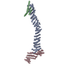
| ||||||||
|---|---|---|---|---|---|---|---|---|---|
| 1 |
| ||||||||
| Unit cell |
|
-Components
| #1: Protein | Mass: 79860.883 Da / Num. of mol.: 1 Source method: isolated from a genetically manipulated source Source: (gene. exp.) Kluyveromyces lactis NRRL Y-1140 (yeast)Strain: ATCC 8585 / CBS 2359 / DSM 70799 / NBRC 1267 / NRRL Y-1140 / WM37 Gene: KLLA0_B05115g / Plasmid: pQLinkH / Production host:  |
|---|---|
| #2: Protein | Mass: 13260.607 Da / Num. of mol.: 1 Source method: isolated from a genetically manipulated source Source: (gene. exp.) Kluyveromyces lactis NRRL Y-1140 (yeast)Strain: ATCC 8585 / CBS 2359 / DSM 70799 / NBRC 1267 / NRRL Y-1140 / WM37 Gene: KLLA0_F06644g / Plasmid: pQLinkH / Production host: |
| #3: Protein | Mass: 36014.375 Da / Num. of mol.: 1 Source method: isolated from a genetically manipulated source Source: (gene. exp.) Kluyveromyces lactis NRRL Y-1140 (yeast)Strain: ATCC 8585 / CBS 2359 / DSM 70799 / NBRC 1267 / NRRL Y-1140 / WM37 Gene: KLLA0_C02695g / Plasmid: pQLinkH / Production host: |
-Experimental details
-Experiment
| Experiment | Method: X-RAY DIFFRACTION / Number of used crystals: 1 |
|---|
- Sample preparation
| Crystal | Density Matthews: 6.88 Å3/Da / Density % sol: 80.48 % / Description: thin rectangular plates, 200 x 200 x 20 um |
|---|---|
| Crystal grow | Temperature: 295 K / Method: vapor diffusion, hanging drop / pH: 7.5 Details: 0.1 M Tris, pH 7.5, 0.1 M NaCl, 8% (w/v) PEG 3350, 10% (v/v) ethylene glycol, 1 mM dithiothreitol. Cryoprotected with 30% (v/v) ethylene glycol |
-Data collection
| Diffraction | Mean temperature: 100 K / Serial crystal experiment: N | |||||||||||||||||||||||||||
|---|---|---|---|---|---|---|---|---|---|---|---|---|---|---|---|---|---|---|---|---|---|---|---|---|---|---|---|---|
| Diffraction source | Source: SYNCHROTRON / Site: NSLS-II / Beamline: 17-ID-1 / Wavelength: 0.97932 Å | |||||||||||||||||||||||||||
| Detector | Type: DECTRIS EIGER X 9M / Detector: PIXEL / Date: Jun 14, 2018 | |||||||||||||||||||||||||||
| Radiation | Monochromator: Si(111) silicon crystal / Protocol: SINGLE WAVELENGTH / Monochromatic (M) / Laue (L): M / Scattering type: x-ray | |||||||||||||||||||||||||||
| Radiation wavelength | Wavelength: 0.97932 Å / Relative weight: 1 | |||||||||||||||||||||||||||
| Reflection | Resolution: 5.726→29.601 Å / Num. obs: 7053 / % possible obs: 74.7 % / Redundancy: 6.8 % / Biso Wilson estimate: 317.29 Å2 / CC1/2: 0.997 / Rmerge(I) obs: 0.115 / Rpim(I) all: 0.07 / Rrim(I) all: 0.135 / Net I/σ(I): 10.7 | |||||||||||||||||||||||||||
| Reflection shell | Diffraction-ID: 1
|
- Processing
| Software |
| ||||||||||||||||||||||||||||||||||||||||||
|---|---|---|---|---|---|---|---|---|---|---|---|---|---|---|---|---|---|---|---|---|---|---|---|---|---|---|---|---|---|---|---|---|---|---|---|---|---|---|---|---|---|---|---|
| Refinement | Method to determine structure: MOLECULAR REPLACEMENT / Resolution: 5.73→29.6 Å / SU ML: 1.06 / Cross valid method: THROUGHOUT / σ(F): 1.36 / Phase error: 40.42 / Stereochemistry target values: ML
| ||||||||||||||||||||||||||||||||||||||||||
| Solvent computation | Shrinkage radii: 0.9 Å / VDW probe radii: 1.11 Å / Solvent model: FLAT BULK SOLVENT MODEL | ||||||||||||||||||||||||||||||||||||||||||
| Refinement step | Cycle: LAST / Resolution: 5.73→29.6 Å
| ||||||||||||||||||||||||||||||||||||||||||
| Refine LS restraints |
| ||||||||||||||||||||||||||||||||||||||||||
| LS refinement shell |
| ||||||||||||||||||||||||||||||||||||||||||
| Refinement TLS params. | Method: refined / Origin x: 9.1493 Å / Origin y: 40.5827 Å / Origin z: -30.9085 Å
| ||||||||||||||||||||||||||||||||||||||||||
| Refinement TLS group | Selection details: all |
