National Institutes of Health/National Institute of General Medical Sciences (NIH/NIGMS)
R01 GM135651
United States
National Institutes of Health/National Institute of General Medical Sciences (NIH/NIGMS)
U24GM129547
United States
Citation
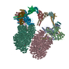
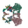
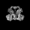
Journal: Sci Adv / Year: 2023 Title: Cryo-EM visualization of DNA-PKcs structural intermediates in NHEJ. Authors: Siyu Chen / Alex Vogt / Linda Lee / Tasmin Naila / Ryan McKeown / Alan E Tomkinson / Susan P Lees-Miller / Yuan He / Abstract: DNA double-strand breaks (DSBs), one of the most cytotoxic forms of DNA damage, can be repaired by the tightly regulated nonhomologous end joining (NHEJ) machinery (Stinson and Loparo and Zhao ). ...DNA double-strand breaks (DSBs), one of the most cytotoxic forms of DNA damage, can be repaired by the tightly regulated nonhomologous end joining (NHEJ) machinery (Stinson and Loparo and Zhao ). Core NHEJ factors form an initial long-range (LR) synaptic complex that transitions into a DNA-PKcs (DNA-dependent protein kinase, catalytic subunit)-free, short-range state to align the DSB ends (Chen ). Using single-particle cryo-electron microscopy, we have visualized three additional key NHEJ complexes representing different transition states, with DNA-PKcs adopting distinct dimeric conformations within each of them. Upon DNA-PKcs autophosphorylation, the LR complex undergoes a substantial conformational change, with both Ku and DNA-PKcs rotating outward to promote DNA break exposure and DNA-PKcs dissociation. We also captured a dimeric state of catalytically inactive DNA-PKcs, which resembles structures of other PIKK (Phosphatidylinositol 3-kinase-related kinase) family kinases, revealing a model of the full regulatory cycle of DNA-PKcs during NHEJ.
History
Deposition
Oct 31, 2022
Deposition site: RCSB / Processing site: RCSB
Revision 1.0
Jun 14, 2023
Provider: repository / Type: Initial release
Revision 1.1
Jun 19, 2024
Group: Data collection / Category: chem_comp_atom / chem_comp_bond
R: unknown region of DNA-PKcs L: DNA-dependent protein kinase catalytic subunit C: DNA-dependent protein kinase catalytic subunit Q: unknown region of DNA-PKcs
Mass: 1720.111 Da / Num. of mol.: 2 / Source method: isolated from a natural source / Source: (natural) Homo sapiens (human) / Cell line: HELA / References: non-specific serine/threonine protein kinase
#2: Protein
DNA-dependentproteinkinasecatalyticsubunit
Mass: 469673.219 Da / Num. of mol.: 2 / Source method: isolated from a natural source / Source: (natural) Homo sapiens (human) / Cell line: HELA / References: UniProt: P78527
-
Experimental details
-
Experiment
Experiment
Method: ELECTRON MICROSCOPY
EM experiment
Aggregation state: PARTICLE / 3D reconstruction method: single particle reconstruction
Cryogen: NITROGEN / Specimen holder model: FEI TITAN KRIOS AUTOGRID HOLDER / Temperature (max): 70 K / Temperature (min): 70 K
Image recording
Average exposure time: 4 sec. / Electron dose: 65 e/Å2 / Film or detector model: GATAN K3 (6k x 4k) / Num. of grids imaged: 1 / Num. of real images: 13132
Image scans
Width: 5760 / Height: 4092
-
Processing
Software
Name: UCSF ChimeraX / Version: 1.4/v9 / Classification: model building / URL: https://www.rbvi.ucsf.edu/chimerax/ / Os: macOS / Type: package
EM software
ID
Name
Version
Category
1
Gautomatch
0.8
particleselection
2
cryoSPARC
2
imageacquisition
4
RELION
3.1.3
CTFcorrection
7
ISOLDE
1.4
modelfitting
9
RELION
3.1.3
initialEulerassignment
10
RELION
3.1.3
finalEulerassignment
11
RELION
3.1.3
classification
12
RELION
3.1.3
3Dreconstruction
13
ISOLDE
1.4
modelrefinement
CTF correction
Type: PHASE FLIPPING AND AMPLITUDE CORRECTION
Particle selection
Num. of particles selected: 1587734
Symmetry
Point symmetry: C2 (2 fold cyclic)
3D reconstruction
Resolution: 5.67 Å / Resolution method: FSC 0.143 CUT-OFF / Num. of particles: 64775 / Algorithm: FOURIER SPACE / Num. of class averages: 1 / Symmetry type: POINT
Atomic model building
Protocol: FLEXIBLE FIT / Space: REAL / Target criteria: CC
Refine LS restraints
Refine-ID
Type
Dev ideal
Number
ELECTRONMICROSCOPY
f_bond_d
0.004
29994
ELECTRONMICROSCOPY
f_angle_d
0.883
40551
ELECTRONMICROSCOPY
f_dihedral_angle_d
7.702
3966
ELECTRONMICROSCOPY
f_chiral_restr
0.048
4604
ELECTRONMICROSCOPY
f_plane_restr
0.006
5161
+
About Yorodumi
-
News
-
Feb 9, 2022. New format data for meta-information of EMDB entries
New format data for meta-information of EMDB entries
Version 3 of the EMDB header file is now the official format.
The previous official version 1.9 will be removed from the archive.
In the structure databanks used in Yorodumi, some data are registered as the other names, "COVID-19 virus" and "2019-nCoV". Here are the details of the virus and the list of structure data.
Jan 31, 2019. EMDB accession codes are about to change! (news from PDBe EMDB page)
EMDB accession codes are about to change! (news from PDBe EMDB page)
The allocation of 4 digits for EMDB accession codes will soon come to an end. Whilst these codes will remain in use, new EMDB accession codes will include an additional digit and will expand incrementally as the available range of codes is exhausted. The current 4-digit format prefixed with “EMD-” (i.e. EMD-XXXX) will advance to a 5-digit format (i.e. EMD-XXXXX), and so on. It is currently estimated that the 4-digit codes will be depleted around Spring 2019, at which point the 5-digit format will come into force.
The EM Navigator/Yorodumi systems omit the EMD- prefix.
Related info.:Q: What is EMD? / ID/Accession-code notation in Yorodumi/EM Navigator
Yorodumi is a browser for structure data from EMDB, PDB, SASBDB, etc.
This page is also the successor to EM Navigator detail page, and also detail information page/front-end page for Omokage search.
The word "yorodu" (or yorozu) is an old Japanese word meaning "ten thousand". "mi" (miru) is to see.
Related info.:EMDB / PDB / SASBDB / Comparison of 3 databanks / Yorodumi Search / Aug 31, 2016. New EM Navigator & Yorodumi / Yorodumi Papers / Jmol/JSmol / Function and homology information / Changes in new EM Navigator and Yorodumi
 Movie
Movie Controller


 Open data
 Basic information
 Components
 Keywords
 Function and homology information
 Homo sapiens (human)
 Authors
 United States, 2items
  Citation
 Structure visualization
 Downloads & links
 Other downloads
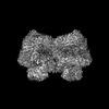


 PDBj
PDBj


 Assembly
 Sample preparation
 Electron microscopy imaging
 FIELD EMISSION GUN / Accelerating voltage: 300 kV / Illumination mode: FLOOD BEAM
 Processing