 Movie
Movie Controller

[English] 日本語
 Yorodumi
- PDB-8czm: Crystal Structure of EcDsbA in a complex with 4-bromo-1H-pyrazole -
+ Open data

- Basic information
| Entry | Database: PDB / ID: 8czm | |||||||||
|---|---|---|---|---|---|---|---|---|---|---|
| Title | Crystal Structure of EcDsbA in a complex with 4-bromo-1H-pyrazole | |||||||||
 Components Components | Thiol:disulfide interchange protein DsbA | |||||||||
 Keywords Keywords | OXIDOREDUCTASE / DISULFIDE OXIDOREDUCTASE / REDOX PROTEIN / Microfrag screening / FBDD / Fragment / ANTIMICROBIAL PROTEIN | |||||||||
| Function / homology |  Function and homology information Function and homology informationprotein disulfide isomerase activity / cellular response to antibiotic / Secretion of toxins / protein-disulfide reductase activity / outer membrane-bounded periplasmic space Similarity search - Function | |||||||||
| Biological species |  | |||||||||
| Method |  X-RAY DIFFRACTION / SYNCHROTRON / MOLECULAR REPLACEMENT / Resolution: 1.8 Å X-RAY DIFFRACTION / SYNCHROTRON / MOLECULAR REPLACEMENT / Resolution: 1.8 Å | |||||||||
 Authors Authors | Whitehouse, R.L. / Ilyichova, O.V. / Taylor, A.J. | |||||||||
| Funding support |  Australia, 2items Australia, 2items
| |||||||||
 Citation Citation | Journal: Rsc Med Chem / Year: 2023 Title: Fragment screening libraries for the identification of protein hot spots and their minimal binding pharmacophores. Authors: Whitehouse, R.L. / Alwan, W.S. / Ilyichova, O.V. / Taylor, A.J. / Chandrashekaran, I.R. / Mohanty, B. / Doak, B.C. / Scanlon, M.J. | |||||||||
| History |
|
- Structure visualization
| Structure viewer | Molecule: MolmilJmol/JSmol |
|---|
- Downloads & links
-Download
| PDBx/mmCIF format | 8czm.cif.gz | 199.4 KB | Display | PDBx/mmCIF format |
|---|---|---|---|---|
| PDB format | pdb8czm.ent.gz | 131.6 KB | Display | PDB format |
| PDBx/mmJSON format | 8czm.json.gz | Tree view | PDBx/mmJSON format | |
| Others |  Other downloads Other downloads |
-Validation report
| Arichive directory | https://data.pdbj.org/pub/pdb/validation_reports/cz/8czmftp://data.pdbj.org/pub/pdb/validation_reports/cz/8czm | HTTPS FTP |
|---|
-Related structure data
| Related structure data | 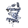 8cxdC 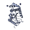 8cxeC 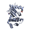 8cznC 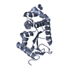 8d10C 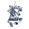 8d11C  8d12C  8dg0C  8dg1C 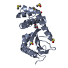 8dg2C  1fvkS S: Starting model for refinement C: citing same article ( |
|---|---|
| Similar structure data |
-Links
 PDBj
PDBj

- Assembly
| Deposited unit | 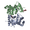
| ||||||||||||||||||||||||||||||||||||||||||||||||||||||||||||||||||||||||||||||||||||||||||||||||||||||||||||||||
|---|---|---|---|---|---|---|---|---|---|---|---|---|---|---|---|---|---|---|---|---|---|---|---|---|---|---|---|---|---|---|---|---|---|---|---|---|---|---|---|---|---|---|---|---|---|---|---|---|---|---|---|---|---|---|---|---|---|---|---|---|---|---|---|---|---|---|---|---|---|---|---|---|---|---|---|---|---|---|---|---|---|---|---|---|---|---|---|---|---|---|---|---|---|---|---|---|---|---|---|---|---|---|---|---|---|---|---|---|---|---|---|---|---|
| 1 | 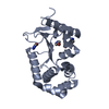
| ||||||||||||||||||||||||||||||||||||||||||||||||||||||||||||||||||||||||||||||||||||||||||||||||||||||||||||||||
| 2 | 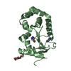
| ||||||||||||||||||||||||||||||||||||||||||||||||||||||||||||||||||||||||||||||||||||||||||||||||||||||||||||||||
| Unit cell |
| ||||||||||||||||||||||||||||||||||||||||||||||||||||||||||||||||||||||||||||||||||||||||||||||||||||||||||||||||
| Noncrystallographic symmetry (NCS) | NCS domain:
NCS domain segments:
|