 Movie
Movie Controller

[English] 日本語
 Yorodumi
- PDB-8czn: Crystal Structure of EcDsbA in a complex with 1H-pyrrole-3-carbox... -
+ Open data

- Basic information
| Entry | Database: PDB / ID: 8czn | |||||||||
|---|---|---|---|---|---|---|---|---|---|---|
| Title | Crystal Structure of EcDsbA in a complex with 1H-pyrrole-3-carboxylic acid | |||||||||
 Components Components | Thiol:disulfide interchange protein DsbA | |||||||||
 Keywords Keywords | OXIDOREDUCTASE / DISULFIDE OXIDOREDUCTASE / REDOX PROTEIN / Microfrag screening / FBDD / Fragment | |||||||||
| Function / homology |  Function and homology information Function and homology informationprotein disulfide isomerase activity / cellular response to antibiotic / Secretion of toxins / protein-disulfide reductase activity / outer membrane-bounded periplasmic space Similarity search - Function | |||||||||
| Biological species |  | |||||||||
| Method |  X-RAY DIFFRACTION / SYNCHROTRON / MOLECULAR REPLACEMENT / Resolution: 1.7 Å X-RAY DIFFRACTION / SYNCHROTRON / MOLECULAR REPLACEMENT / Resolution: 1.7 Å | |||||||||
 Authors Authors | Whitehouse, R.L. / Ilyichova, O.V. / Taylor, A.J. | |||||||||
| Funding support |  Australia, 2items Australia, 2items
| |||||||||
 Citation Citation | Journal: Rsc Med Chem / Year: 2023 Title: Fragment screening libraries for the identification of protein hot spots and their minimal binding pharmacophores. Authors: Whitehouse, R.L. / Alwan, W.S. / Ilyichova, O.V. / Taylor, A.J. / Chandrashekaran, I.R. / Mohanty, B. / Doak, B.C. / Scanlon, M.J. | |||||||||
| History |
|
- Structure visualization
| Structure viewer | Molecule: MolmilJmol/JSmol |
|---|
- Downloads & links
-Download
| PDBx/mmCIF format | 8czn.cif.gz | 205.7 KB | Display | PDBx/mmCIF format |
|---|---|---|---|---|
| PDB format | pdb8czn.ent.gz | 136.2 KB | Display | PDB format |
| PDBx/mmJSON format | 8czn.json.gz | Tree view | PDBx/mmJSON format | |
| Others |  Other downloads Other downloads |
-Validation report
| Arichive directory | https://data.pdbj.org/pub/pdb/validation_reports/cz/8cznftp://data.pdbj.org/pub/pdb/validation_reports/cz/8czn | HTTPS FTP |
|---|
-Related structure data
| Related structure data |  8cxdC  8cxeC  8czmC  8d10C  8d11C  8d12C  8dg0C  8dg1C 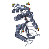 8dg2C  1fvkS S: Starting model for refinement C: citing same article ( |
|---|---|
| Similar structure data |
-Links
 PDBj
PDBj

- Assembly
| Deposited unit | 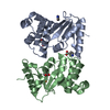
| ||||||||||||||||||||||||||||||||||||||||||||||||||||||||||||||||||||||||
|---|---|---|---|---|---|---|---|---|---|---|---|---|---|---|---|---|---|---|---|---|---|---|---|---|---|---|---|---|---|---|---|---|---|---|---|---|---|---|---|---|---|---|---|---|---|---|---|---|---|---|---|---|---|---|---|---|---|---|---|---|---|---|---|---|---|---|---|---|---|---|---|---|---|
| 1 | 
| ||||||||||||||||||||||||||||||||||||||||||||||||||||||||||||||||||||||||
| 2 | 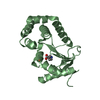
| ||||||||||||||||||||||||||||||||||||||||||||||||||||||||||||||||||||||||
| Unit cell |
| ||||||||||||||||||||||||||||||||||||||||||||||||||||||||||||||||||||||||
| Noncrystallographic symmetry (NCS) | NCS domain:
NCS domain segments:
NCS oper: (Code: givenMatrix: (-0.965607807855, 0.0637850787006, 0.252057582994), (-0.251770538593, 0.0126309586466, -0.967704528655), (-0.0649088384265, -0.997883721993, 0.00386265132877)Vector: 36. ...NCS oper: (Code: given Matrix: (-0.965607807855, 0.0637850787006, 0.252057582994), Vector: |
-Components
| #1: Protein | Mass: 21155.025 Da / Num. of mol.: 2 Source method: isolated from a genetically manipulated source Source: (gene. exp.) #2: Chemical | ChemComp-PKN /   Mass: 111.099 Da / Num. of mol.: 4 / Source method: obtained synthetically / Formula: C5H5NO2 / Feature type: SUBJECT OF INVESTIGATION Mass: 111.099 Da / Num. of mol.: 4 / Source method: obtained synthetically / Formula: C5H5NO2 / Feature type: SUBJECT OF INVESTIGATION#3: Chemical | ChemComp-CU / |   Mass: 63.546 Da / Num. of mol.: 1 / Source method: obtained synthetically / Formula: Cu Mass: 63.546 Da / Num. of mol.: 1 / Source method: obtained synthetically / Formula: Cu#4: Water | ChemComp-HOH / |  Mass: 18.015 Da / Num. of mol.: 360 / Source method: isolated from a natural source / Formula: H2O Mass: 18.015 Da / Num. of mol.: 360 / Source method: isolated from a natural source / Formula: H2OHas ligand of interest | Y | Has protein modification | Y | |
|---|
-Experimental details
-Experiment
| Experiment | Method: X-RAY DIFFRACTION / Number of used crystals: 1 |
|---|
- Sample preparation
| Crystal | Density Matthews: 2.8 Å3/Da / Density % sol: 56.13 % |
|---|---|
| Crystal grow | Temperature: 293.15 K / Method: vapor diffusion, hanging drop / pH: 6.1 Details: 11-13 % PEG 8000, 5-7.5% GLYCEROL, 100MM NA CACODYLATE PH6.1, 1MM CuCl2 |
-Data collection
| Diffraction | Mean temperature: 100 K / Serial crystal experiment: N |
|---|---|
| Diffraction source | Source: SYNCHROTRON / Site: Australian Synchrotron / Beamline: MX2 / Wavelength: 0.95365 Å |
| Detector | Type: DECTRIS EIGER X 16M / Detector: PIXEL / Date: Sep 24, 2019 |
| Radiation | Protocol: SINGLE WAVELENGTH / Monochromatic (M) / Laue (L): M / Scattering type: x-ray |
| Radiation wavelength | Wavelength: 0.95365 Å / Relative weight: 1 |
| Reflection | Resolution: 1.59→48.14 Å / Num. obs: 62171 / % possible obs: 99.1 % / Redundancy: 6.7 % / Biso Wilson estimate: 22.88 Å2 / CC1/2: 0.999 / Rmerge(I) obs: 0.084 / Rpim(I) all: 0.053 / Rrim(I) all: 0.1 / Χ2: 0.5 / Net I/σ(I): 10.6 |
| Reflection shell | Resolution: 1.59→1.61 Å / Redundancy: 5.9 % / Rmerge(I) obs: 1.726 / Mean I/σ(I) obs: 0.8 / Num. unique obs: 2545 / CC1/2: 0.369 / Rpim(I) all: 0.821 / Χ2: 0.369 / % possible all: 82.5 |
- Processing
| Software |
| |||||||||||||||||||||||||||||||||||||||||||||||||
|---|---|---|---|---|---|---|---|---|---|---|---|---|---|---|---|---|---|---|---|---|---|---|---|---|---|---|---|---|---|---|---|---|---|---|---|---|---|---|---|---|---|---|---|---|---|---|---|---|---|---|
| Refinement | Method to determine structure: MOLECULAR REPLACEMENT Starting model: 1FVK Resolution: 1.7→31.6 Å / SU ML: 0.2367 / Cross valid method: FREE R-VALUE / σ(F): 1.38 / Phase error: 23.3131 Stereochemistry target values: GeoStd + Monomer Library + CDL v1.2
| |||||||||||||||||||||||||||||||||||||||||||||||||
| Solvent computation | Shrinkage radii: 0.9 Å / VDW probe radii: 1.11 Å / Solvent model: FLAT BULK SOLVENT MODEL | |||||||||||||||||||||||||||||||||||||||||||||||||
| Displacement parameters | Biso mean: 27.34 Å2 | |||||||||||||||||||||||||||||||||||||||||||||||||
| Refinement step | Cycle: LAST / Resolution: 1.7→31.6 Å
| |||||||||||||||||||||||||||||||||||||||||||||||||
| Refine LS restraints |
| |||||||||||||||||||||||||||||||||||||||||||||||||
| Refine LS restraints NCS | Type: Torsion NCS / Rms dev position: 1.0782598151 Å | |||||||||||||||||||||||||||||||||||||||||||||||||
| LS refinement shell |
| |||||||||||||||||||||||||||||||||||||||||||||||||
| Refinement TLS params. | Method: refined / Origin x: 19.9043326833 Å / Origin y: 0.102637454795 Å / Origin z: 14.2727659789 Å
| |||||||||||||||||||||||||||||||||||||||||||||||||
| Refinement TLS group | Selection details: all |
