ムービー
ムービー コントローラー


+ データを開く

- 基本情報
基本情報
| 登録情報 | データベース: PDB / ID: 8cqb | |||||||||
|---|---|---|---|---|---|---|---|---|---|---|
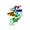
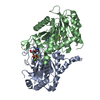
| タイトル | Cryo-EM structure of the human GBP1 dimer bound to GDP-AlF3 | |||||||||
 要素 要素 | Guanylate-binding protein 1 | |||||||||
 キーワード キーワード | IMMUNE SYSTEM / Cell-autonomous immunity / intracellular pathogens / GTPase | |||||||||
| 機能・相同性 |  機能・相同性情報 機能・相同性情報GDP phosphatase activity / non-canonical inflammasome complex assembly / negative regulation of substrate adhesion-dependent cell spreading / protein localization to vacuole / positive regulation of pyroptotic inflammatory response / cytolysis in another organism / symbiont cell surface / vesicle membrane / negative regulation of interleukin-2 production / spectrin binding ...GDP phosphatase activity / non-canonical inflammasome complex assembly / negative regulation of substrate adhesion-dependent cell spreading / protein localization to vacuole / positive regulation of pyroptotic inflammatory response / cytolysis in another organism / symbiont cell surface / vesicle membrane / negative regulation of interleukin-2 production / spectrin binding / defense response to protozoan / cytokine binding / cellular response to interleukin-1 / negative regulation of T cell receptor signaling pathway / negative regulation of protein localization to plasma membrane / regulation of protein localization to plasma membrane / 加水分解酵素; 酸無水物に作用; リン含有酸無水物に作用 / regulation of calcium-mediated signaling / cytoplasmic vesicle membrane / lipopolysaccharide binding / Hsp90 protein binding / negative regulation of ERK1 and ERK2 cascade / cellular response to type II interferon / cellular response to tumor necrosis factor / Interferon gamma signaling / GDP binding / actin cytoskeleton / actin binding / G protein activity / cytoplasmic vesicle / defense response to virus / 加水分解酵素; 酸無水物に作用; GTPに作用・細胞または細胞小器官の運動に関与 / defense response to bacterium / Golgi membrane / innate immune response / GTPase activity / GTP binding / enzyme binding / Golgi apparatus / protein homodimerization activity / extracellular region / identical protein binding / plasma membrane / cytoplasm / cytosol 類似検索 - 分子機能 | |||||||||
| 生物種 |  Homo sapiens (ヒト) Homo sapiens (ヒト) | |||||||||
| 手法 | 電子顕微鏡法 / 単粒子再構成法 / クライオ電子顕微鏡法 / 解像度: 3.7 Å | |||||||||
 データ登録者 データ登録者 | Kuhm, T.I. / Jakobi, A.J. | |||||||||
| 資金援助 | European Union,  オランダ, 2件 オランダ, 2件
| |||||||||
 引用 引用 | ジャーナル: Nat Struct Mol Biol / 年: 2025 タイトル: Structural basis of antimicrobial membrane coat assembly by human GBP1. 著者: Tanja Kuhm / Clémence Taisne / Cecilia de Agrela Pinto / Luca Gross / Evdokia A Giannopoulou / Stefan T Huber / Els Pardon / Jan Steyaert / Sander J Tans / Arjen J Jakobi /  要旨: Guanylate-binding proteins (GBPs) are interferon-inducible guanosine triphosphate hydrolases (GTPases) mediating host defense against intracellular pathogens. Their antimicrobial activity hinges on ...Guanylate-binding proteins (GBPs) are interferon-inducible guanosine triphosphate hydrolases (GTPases) mediating host defense against intracellular pathogens. Their antimicrobial activity hinges on their ability to self-associate and coat pathogen-associated compartments or cytosolic bacteria. Coat formation depends on GTPase activity but how nucleotide binding and hydrolysis prime coat formation remains unclear. Here, we report the cryo-electron microscopy structure of the full-length human GBP1 dimer in its guanine nucleotide-bound state and describe the molecular ultrastructure of the GBP1 coat on liposomes and bacterial lipopolysaccharide membranes. Conformational changes of the middle and GTPase effector domains expose the isoprenylated C terminus for membrane association. The α-helical middle domains form a parallel, crossover arrangement essential for coat formation and position the extended effector domain for intercalation into the lipopolysaccharide layer of gram-negative membranes. Nucleotide binding and hydrolysis create oligomeric scaffolds with contractile abilities that promote membrane extrusion and fragmentation. Our data offer a structural and mechanistic framework for understanding GBP1 effector functions in intracellular immunity. | |||||||||
| 履歴 |
|
- 構造の表示
構造の表示
| 構造ビューア | 分子: MolmilJmol/JSmol |
|---|
- ダウンロードとリンク
-ダウンロード
| PDBx/mmCIF形式 | 8cqb.cif.gz | 181.9 KB | 表示 | PDBx/mmCIF形式 |
|---|---|---|---|---|
| PDB形式 | pdb8cqb.ent.gz | 142.8 KB | 表示 | PDB形式 |
| PDBx/mmJSON形式 | 8cqb.json.gz | ツリー表示 | PDBx/mmJSON形式 | |
| その他 |  その他のダウンロード その他のダウンロード |
-検証レポート
| アーカイブディレクトリ | https://data.pdbj.org/pub/pdb/validation_reports/cq/8cqbftp://data.pdbj.org/pub/pdb/validation_reports/cq/8cqb | HTTPS FTP |
|---|
-関連構造データ

-リンク
 PDBj
PDBj

- 集合体
集合体
| 登録構造単位 | 
|
|---|---|
| 1 |
|
-要素
| #1: タンパク質 | 分子量: 68021.617 Da / 分子数: 2 / 由来タイプ: 組換発現 / 由来: (組換発現) Homo sapiens (ヒト) / 遺伝子: GBP1 / 発現宿主:  参照: UniProt: P32455, 加水分解酵素; 酸無水物に作用; GTPに作用・細胞または細胞小器官の運動に関与 #2: 化合物 |   タイプ: RNA linking / 分子量: 443.201 Da / 分子数: 2 / 由来タイプ: 合成 / 式: C10H15N5O11P2 / タイプ: SUBJECT OF INVESTIGATION / コメント: GDP, エネルギー貯蔵分子*YM タイプ: RNA linking / 分子量: 443.201 Da / 分子数: 2 / 由来タイプ: 合成 / 式: C10H15N5O11P2 / タイプ: SUBJECT OF INVESTIGATION / コメント: GDP, エネルギー貯蔵分子*YM#3: 化合物 |   分子量: 83.977 Da / 分子数: 2 / 由来タイプ: 合成 / 式: AlF3 / タイプ: SUBJECT OF INVESTIGATION 分子量: 83.977 Da / 分子数: 2 / 由来タイプ: 合成 / 式: AlF3 / タイプ: SUBJECT OF INVESTIGATION#4: 化合物 |   分子量: 24.305 Da / 分子数: 2 / 由来タイプ: 合成 / 式: Mg / タイプ: SUBJECT OF INVESTIGATION 分子量: 24.305 Da / 分子数: 2 / 由来タイプ: 合成 / 式: Mg / タイプ: SUBJECT OF INVESTIGATION研究の焦点であるリガンドがあるか | Y | Has protein modification | N | |
|---|
-実験情報
-実験
| 実験 | 手法: 電子顕微鏡法 |
|---|---|
| EM実験 | 試料の集合状態: PARTICLE / 3次元再構成法: 単粒子再構成法 |
- 試料調製
試料調製
| 構成要素 | 名称: Homodimeric complex of GBP1 stabilised by GDP-AlF3 / タイプ: COMPLEX / Entity ID: #1 / 由来: RECOMBINANT | ||||||||||||||||||||||||||||||||||||||||
|---|---|---|---|---|---|---|---|---|---|---|---|---|---|---|---|---|---|---|---|---|---|---|---|---|---|---|---|---|---|---|---|---|---|---|---|---|---|---|---|---|---|
| 分子量 | 値: 0.134 MDa / 実験値: YES | ||||||||||||||||||||||||||||||||||||||||
| 由来(天然) | 生物種: Homo sapiens (ヒト) | ||||||||||||||||||||||||||||||||||||||||
| 由来(組換発現) | 生物種: | ||||||||||||||||||||||||||||||||||||||||
| 緩衝液 | pH: 7.4 詳細: 5u mM HEPES pH7.4, 150 mM NaCl, 1 mM DTT, 200 uM GDP, 10 mM NaF, 300 uM AlCl3, 5 mM MgCl2 | ||||||||||||||||||||||||||||||||||||||||
| 緩衝液成分 |
| ||||||||||||||||||||||||||||||||||||||||
| 試料 | 濃度: 0.7 mg/ml / 包埋: NO / シャドウイング: NO / 染色: NO / 凍結: YES | ||||||||||||||||||||||||||||||||||||||||
| 試料支持 | グリッドの材料: COPPER / グリッドのサイズ: 300 divisions/in. / グリッドのタイプ: Quantifoil | ||||||||||||||||||||||||||||||||||||||||
| 急速凍結 | 装置: LEICA EM GP / 凍結剤: ETHANE / 湿度: 99 % / 凍結前の試料温度: 22 K |
- 電子顕微鏡撮影
| 実験機器 |  モデル: Titan Krios / 画像提供: FEI Company |
|---|---|
| 顕微鏡 | モデル: FEI TITAN KRIOS |
| 電子銃 | 電子線源:  FIELD EMISSION GUN / 加速電圧: 300 kV / 照射モード: FLOOD BEAM FIELD EMISSION GUN / 加速電圧: 300 kV / 照射モード: FLOOD BEAM |
| 電子レンズ | モード: BRIGHT FIELD / 倍率(公称値): 105000 X / 最大 デフォーカス(公称値): 2200 nm / 最小 デフォーカス(公称値): 600 nm / Cs: 2.7 mm / アライメント法: COMA FREE |
| 試料ホルダ | 凍結剤: NITROGEN 試料ホルダーモデル: FEI TITAN KRIOS AUTOGRID HOLDER |
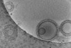
| 撮影 | 電子線照射量: 60 e/Å2 フィルム・検出器のモデル: GATAN K3 BIOQUANTUM (6k x 4k) 撮影したグリッド数: 2 / 実像数: 5214 |
| 電子光学装置 | エネルギーフィルター名称: GIF Bioquantum / エネルギーフィルタースリット幅: 20 eV |
| 画像スキャン | 横: 5760 / 縦: 4092 |
- 解析
解析
| ソフトウェア | 名称: UCSF ChimeraX / バージョン: 1.3/v9 / 分類: モデル構築 / URL: https://www.rbvi.ucsf.edu/chimerax/ / Os: macOS / タイプ: package | ||||||||||||||||||||||||||||||||
|---|---|---|---|---|---|---|---|---|---|---|---|---|---|---|---|---|---|---|---|---|---|---|---|---|---|---|---|---|---|---|---|---|---|
| EMソフトウェア |
| ||||||||||||||||||||||||||||||||
| CTF補正 | タイプ: PHASE FLIPPING AND AMPLITUDE CORRECTION | ||||||||||||||||||||||||||||||||
| 粒子像の選択 | 選択した粒子像数: 447651 詳細: 447,651 particles were used to perform ab initio reconstruction to generate five different models. Three classes were selected for heterogeneous refinements without imposing symmetry. A ...詳細: 447,651 particles were used to perform ab initio reconstruction to generate five different models. Three classes were selected for heterogeneous refinements without imposing symmetry. A single class with 147095 particles was used for non-uniform refinement. | ||||||||||||||||||||||||||||||||
| 対称性 | 点対称性: C1 (非対称) | ||||||||||||||||||||||||||||||||
| 3次元再構成 | 解像度: 3.7 Å / 解像度の算出法: FSC 0.143 CUT-OFF / 粒子像の数: 147095 / 対称性のタイプ: POINT | ||||||||||||||||||||||||||||||||
| 原子モデル構築 | プロトコル: OTHER / 空間: RECIPROCAL / Target criteria: Real-space cross correlation | ||||||||||||||||||||||||||||||||
| 原子モデル構築 | 3D fitting-ID: 1 / Source name: PDB / タイプ: experimental model
|