 Movie
Movie Controller

[English] 日本語
 Yorodumi
- PDB-8aas: Crystal structure of the Pyrococcus abyssi RPA trimerization core... -
+ Open data

- Basic information
| Entry | Database: PDB / ID: 8aas | |||||||||
|---|---|---|---|---|---|---|---|---|---|---|
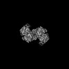
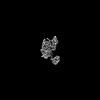
| Title | Crystal structure of the Pyrococcus abyssi RPA trimerization core bound to poly-dT20 ssDNA | |||||||||
 Components Components |
| |||||||||
 Keywords Keywords | DNA BINDING PROTEIN / Replication protein A / ssDNA-Binding protein | |||||||||
| Function / homology |  Function and homology information Function and homology informationresponse to ionizing radiation / double-strand break repair via homologous recombination / DNA binding / metal ion binding Similarity search - Function | |||||||||
| Biological species |   Pyrococcus abyssi GE5 (archaea) Pyrococcus abyssi GE5 (archaea)synthetic construct (others) | |||||||||
| Method |  X-RAY DIFFRACTION / SYNCHROTRON / MOLECULAR REPLACEMENT / Resolution: 3.2 Å X-RAY DIFFRACTION / SYNCHROTRON / MOLECULAR REPLACEMENT / Resolution: 3.2 Å | |||||||||
 Authors Authors | Madru, C. / Legrand, P. / Sauguet, L. | |||||||||
| Funding support |  France, 2items France, 2items
| |||||||||
 Citation Citation | Journal: Nat Commun / Year: 2023 Title: DNA-binding mechanism and evolution of replication protein A. Authors: Clément Madru / Markel Martínez-Carranza / Sébastien Laurent / Alessandra C Alberti / Maelenn Chevreuil / Bertrand Raynal / Ahmed Haouz / Rémy A Le Meur / Marc Delarue / Ghislaine ...Authors: Clément Madru / Markel Martínez-Carranza / Sébastien Laurent / Alessandra C Alberti / Maelenn Chevreuil / Bertrand Raynal / Ahmed Haouz / Rémy A Le Meur / Marc Delarue / Ghislaine Henneke / Didier Flament / Mart Krupovic / Pierre Legrand / Ludovic Sauguet / Abstract: Replication Protein A (RPA) is a heterotrimeric single stranded DNA-binding protein with essential roles in DNA replication, recombination and repair. Little is known about the structure of RPA in ...Replication Protein A (RPA) is a heterotrimeric single stranded DNA-binding protein with essential roles in DNA replication, recombination and repair. Little is known about the structure of RPA in Archaea, the third domain of life. By using an integrative structural, biochemical and biophysical approach, we extensively characterize RPA from Pyrococcus abyssi in the presence and absence of DNA. The obtained X-ray and cryo-EM structures reveal that the trimerization core and interactions promoting RPA clustering on ssDNA are shared between archaea and eukaryotes. However, we also identified a helical domain named AROD (Acidic Rpa1 OB-binding Domain), and showed that, in Archaea, RPA forms an unanticipated tetrameric supercomplex in the absence of DNA. The four RPA molecules clustered within the tetramer could efficiently coat and protect stretches of ssDNA created by the advancing replisome. Finally, our results provide insights into the evolution of this primordial replication factor in eukaryotes. | |||||||||
| History |
|
- Structure visualization
| Structure viewer | Molecule: MolmilJmol/JSmol |
|---|
- Downloads & links
-Download
| PDBx/mmCIF format | 8aas.cif.gz | 224.7 KB | Display | PDBx/mmCIF format |
|---|---|---|---|---|
| PDB format | pdb8aas.ent.gz | 178.5 KB | Display | PDB format |
| PDBx/mmJSON format | 8aas.json.gz | Tree view | PDBx/mmJSON format | |
| Others |  Other downloads Other downloads |
-Validation report
| Arichive directory | https://data.pdbj.org/pub/pdb/validation_reports/aa/8aasftp://data.pdbj.org/pub/pdb/validation_reports/aa/8aas | HTTPS FTP |
|---|
-Related structure data
| Related structure data |  8aa9C 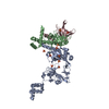 8aajC 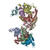 8c5yC 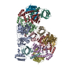 8c5zC 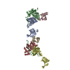 8oejC 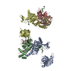 8oelC C: citing same article ( |
|---|---|
| Similar structure data |

-Links
 PDBj
PDBj


- Assembly
| Deposited unit | 
| ||||||||
|---|---|---|---|---|---|---|---|---|---|
| 1 |
| ||||||||
| Unit cell |
|
-Components
-Protein , 3 types, 3 molecules ABC
| #1: Protein | Mass: 33756.676 Da / Num. of mol.: 1 Source method: isolated from a genetically manipulated source Source: (gene. exp.) Pyrococcus abyssi GE5 (archaea) / Strain: GE5 / Orsay / Gene: PAB2163 / Production host:  |
|---|---|
| #2: Protein | Mass: 21277.865 Da / Num. of mol.: 1 Source method: isolated from a genetically manipulated source Details: Insertion of SER after first M from reference sequence NCBI Reference Sequence: WP_048146526.1 to improve expression Source: (gene. exp.) Pyrococcus abyssi GE5 (archaea) / Strain: GE5 / Orsay / Gene: PAB2165 / Production host: |
| #3: Protein | Mass: 14008.925 Da / Num. of mol.: 1 Source method: isolated from a genetically manipulated source Source: (gene. exp.) Pyrococcus abyssi GE5 (archaea) / Strain: GE5 / Orsay / Gene: PAB2164 / Production host: |
-DNA chain , 1 types, 1 molecules F
| #4: DNA chain | Mass: 6038.899 Da / Num. of mol.: 1 / Source method: obtained synthetically / Source: (synth.) synthetic construct (others) |
|---|
-Non-polymers , 2 types, 6 molecules 

| #5: Chemical | ChemComp-ZN /  Mass: 65.409 Da / Num. of mol.: 1 / Source method: obtained synthetically / Formula: Zn / Feature type: SUBJECT OF INVESTIGATION Mass: 65.409 Da / Num. of mol.: 1 / Source method: obtained synthetically / Formula: Zn / Feature type: SUBJECT OF INVESTIGATION |
|---|---|
| #6: Chemical | ChemComp-CA /  Mass: 40.078 Da / Num. of mol.: 5 / Source method: obtained synthetically / Formula: Ca Mass: 40.078 Da / Num. of mol.: 5 / Source method: obtained synthetically / Formula: Ca |
-Details
| Has ligand of interest | Y |
|---|
-Experimental details
-Experiment
| Experiment | Method: X-RAY DIFFRACTION / Number of used crystals: 1 |
|---|
- Sample preparation
| Crystal | Density Matthews: 2.4 Å3/Da / Density % sol: 48.82 % |
|---|---|
| Crystal grow | Temperature: 277 K / Method: vapor diffusion, hanging drop / pH: 8 Details: 10% w/v PEG 8K 0.1M imidazole pH 8 0.2 M calcium acetate |
-Data collection
| Diffraction | Mean temperature: 100 K / Serial crystal experiment: N |
|---|---|
| Diffraction source | Source: SYNCHROTRON / Site: SOLEIL / Beamline: PROXIMA 1 / Wavelength: 0.978565 Å |
| Detector | Type: DECTRIS EIGER X 16M / Detector: PIXEL / Date: Dec 9, 2021 / Details: KB Mirrors |
| Radiation | Monochromator: Si(111) / Protocol: SINGLE WAVELENGTH / Monochromatic (M) / Laue (L): M / Scattering type: x-ray |
| Radiation wavelength | Wavelength: 0.978565 Å / Relative weight: 1 |
| Reflection | Resolution: 3.2→48.26 Å / Num. obs: 10041 / % possible obs: 80.7 % / Redundancy: 27.2 % / Biso Wilson estimate: 163.2 Å2 / CC1/2: 1 / Rmerge(I) obs: 0.067 / Rpim(I) all: 0.013 / Rrim(I) all: 0.068 / Net I/σ(I): 27.1 |
| Reflection shell | Resolution: 3.2→3.28 Å / Redundancy: 26.8 % / Rmerge(I) obs: 4.189 / Mean I/σ(I) obs: 1 / Num. unique obs: 204 / CC1/2: 0.454 / Rpim(I) all: 0.816 / Rrim(I) all: 4.27 / % possible all: 21.2 |
- Processing
| Software |
| ||||||||||||||||||||||||||||||||||||||||||||||||||||||||||||||||||||||||||||||||||||||||||||||||||||
|---|---|---|---|---|---|---|---|---|---|---|---|---|---|---|---|---|---|---|---|---|---|---|---|---|---|---|---|---|---|---|---|---|---|---|---|---|---|---|---|---|---|---|---|---|---|---|---|---|---|---|---|---|---|---|---|---|---|---|---|---|---|---|---|---|---|---|---|---|---|---|---|---|---|---|---|---|---|---|---|---|---|---|---|---|---|---|---|---|---|---|---|---|---|---|---|---|---|---|---|---|---|
| Refinement | Method to determine structure: MOLECULAR REPLACEMENT Starting model: Working model of the full apo-RPA Resolution: 3.2→48.26 Å / Cor.coef. Fo:Fc: 0.934 / Cor.coef. Fo:Fc free: 0.926 / Cross valid method: THROUGHOUT / SU Rfree Blow DPI: 0.634
| ||||||||||||||||||||||||||||||||||||||||||||||||||||||||||||||||||||||||||||||||||||||||||||||||||||
| Displacement parameters | Biso mean: 173.43 Å2
| ||||||||||||||||||||||||||||||||||||||||||||||||||||||||||||||||||||||||||||||||||||||||||||||||||||
| Refine analyze | Luzzati coordinate error obs: 0.59 Å | ||||||||||||||||||||||||||||||||||||||||||||||||||||||||||||||||||||||||||||||||||||||||||||||||||||
| Refinement step | Cycle: LAST / Resolution: 3.2→48.26 Å
| ||||||||||||||||||||||||||||||||||||||||||||||||||||||||||||||||||||||||||||||||||||||||||||||||||||
| Refine LS restraints |
| ||||||||||||||||||||||||||||||||||||||||||||||||||||||||||||||||||||||||||||||||||||||||||||||||||||
| LS refinement shell | Resolution: 3.2→3.35 Å
| ||||||||||||||||||||||||||||||||||||||||||||||||||||||||||||||||||||||||||||||||||||||||||||||||||||
| Refinement TLS params. | Refine-ID: X-RAY DIFFRACTION
| ||||||||||||||||||||||||||||||||||||||||||||||||||||||||||||||||||||||||||||||||||||||||||||||||||||
| Refinement TLS group |
|
