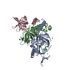
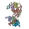
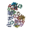
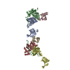
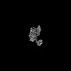
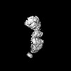
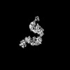
- PDB-8aa9: Crystal structure of the Rpa1 AROD-OB-1 domains -
+
Open data
ID or keywords:
Loading...
-
Basic information
Entry
Database: PDB / ID: 8aa9
Title
Crystal structure of the Rpa1 AROD-OB-1 domains
Components
Replication factor A
Keywords
DNA BINDING PROTEIN / Replication protein A / ssDNA-Binding protein
Function / homology
Function and homology information
response to ionizing radiation / double-strand break repair via homologous recombination / DNA binding / metal ion binding Similarity search - Function
Journal: Nat Commun / Year: 2023 Title: DNA-binding mechanism and evolution of replication protein A. Authors: Clément Madru / Markel Martínez-Carranza / Sébastien Laurent / Alessandra C Alberti / Maelenn Chevreuil / Bertrand Raynal / Ahmed Haouz / Rémy A Le Meur / Marc Delarue / Ghislaine ...Authors: Clément Madru / Markel Martínez-Carranza / Sébastien Laurent / Alessandra C Alberti / Maelenn Chevreuil / Bertrand Raynal / Ahmed Haouz / Rémy A Le Meur / Marc Delarue / Ghislaine Henneke / Didier Flament / Mart Krupovic / Pierre Legrand / Ludovic Sauguet / Abstract: Replication Protein A (RPA) is a heterotrimeric single stranded DNA-binding protein with essential roles in DNA replication, recombination and repair. Little is known about the structure of RPA in ...Replication Protein A (RPA) is a heterotrimeric single stranded DNA-binding protein with essential roles in DNA replication, recombination and repair. Little is known about the structure of RPA in Archaea, the third domain of life. By using an integrative structural, biochemical and biophysical approach, we extensively characterize RPA from Pyrococcus abyssi in the presence and absence of DNA. The obtained X-ray and cryo-EM structures reveal that the trimerization core and interactions promoting RPA clustering on ssDNA are shared between archaea and eukaryotes. However, we also identified a helical domain named AROD (Acidic Rpa1 OB-binding Domain), and showed that, in Archaea, RPA forms an unanticipated tetrameric supercomplex in the absence of DNA. The four RPA molecules clustered within the tetramer could efficiently coat and protect stretches of ssDNA created by the advancing replisome. Finally, our results provide insights into the evolution of this primordial replication factor in eukaryotes.
Method to determine structure: MOLECULAR REPLACEMENT Starting model: Working model of the full RPA Resolution: 1.8→32.25 Å / Cor.coef. Fo:Fc: 0.956 / Cor.coef. Fo:Fc free: 0.95 / SU R Cruickshank DPI: 0.138 / Cross valid method: THROUGHOUT / SU R Blow DPI: 0.142 / SU Rfree Blow DPI: 0.124 / SU Rfree Cruickshank DPI: 0.123
Rfactor
Num. reflection
% reflection
Selection details
Rfree
0.2284
1857
4.92 %
RANDOM
Rwork
0.2086
-
-
-
obs
0.2095
37716
92.4 %
-
Displacement parameters
Biso mean: 39.74 Å2
Baniso -1
Baniso -2
Baniso -3
1-
-0.0597 Å2
0 Å2
0.4556 Å2
2-
-
-1.2775 Å2
0 Å2
3-
-
-
1.3372 Å2
Refine analyze
Luzzati coordinate error obs: 0.26 Å
Refinement step
Cycle: LAST / Resolution: 1.8→32.25 Å
Protein
Nucleic acid
Ligand
Solvent
Total
Num. atoms
2860
0
8
290
3158
Refine LS restraints
Refine-ID
Type
Dev ideal
Number
Restraint function
Weight
X-RAY DIFFRACTION
t_bond_d
0.008
2967
HARMONIC
2
X-RAY DIFFRACTION
t_angle_deg
0.95
4012
HARMONIC
2
X-RAY DIFFRACTION
t_dihedral_angle_d
1127
SINUSOIDAL
2
X-RAY DIFFRACTION
t_gen_planes
501
HARMONIC
5
X-RAY DIFFRACTION
t_it
2967
HARMONIC
10
X-RAY DIFFRACTION
t_chiral_improper_torsion
384
SEMIHARMONIC
5
X-RAY DIFFRACTION
t_sum_occupancies
4
HARMONIC
1
X-RAY DIFFRACTION
t_ideal_dist_contact
2914
SEMIHARMONIC
4
X-RAY DIFFRACTION
t_omega_torsion
3.49
X-RAY DIFFRACTION
t_other_torsion
14.32
LS refinement shell
Resolution: 1.8→1.83 Å
Rfactor
Num. reflection
% reflection
Rfree
0.3423
26
3.4 %
Rwork
0.3088
729
-
obs
-
-
36.75 %
Refinement TLS params.
Origin x: 27.171 Å / Origin y: 45.6439 Å / Origin z: 0.8697 Å
11
12
13
21
22
23
31
32
33
T
-0.001 Å2
0.0089 Å2
-0.0009 Å2
-
-0.0416 Å2
0.0133 Å2
-
-
0.0111 Å2
L
0.5985 °2
-0.1175 °2
-0.1837 °2
-
0.0133 °2
0.0567 °2
-
-
-0.0148 °2
S
0.0015 Å °
-0.0447 Å °
-0.0215 Å °
-0.0447 Å °
-0.0125 Å °
0.013 Å °
-0.0215 Å °
0.013 Å °
0.011 Å °
Refinement TLS group
ID
Refine-ID
Refine TLS-ID
Selection details
Auth asym-ID
Auth seq-ID
1
X-RAY DIFFRACTION
1
{ *|* }
A
3 - 180
2
X-RAY DIFFRACTION
1
{ *|* }
A
201
3
X-RAY DIFFRACTION
1
{ *|* }
B
2 - 180
4
X-RAY DIFFRACTION
1
{ *|* }
C
1
5
X-RAY DIFFRACTION
1
{ *|* }
S
1 - 290
+
About Yorodumi
-
News
-
Feb 9, 2022. New format data for meta-information of EMDB entries
New format data for meta-information of EMDB entries
Version 3 of the EMDB header file is now the official format.
The previous official version 1.9 will be removed from the archive.
In the structure databanks used in Yorodumi, some data are registered as the other names, "COVID-19 virus" and "2019-nCoV". Here are the details of the virus and the list of structure data.
Jan 31, 2019. EMDB accession codes are about to change! (news from PDBe EMDB page)
EMDB accession codes are about to change! (news from PDBe EMDB page)
The allocation of 4 digits for EMDB accession codes will soon come to an end. Whilst these codes will remain in use, new EMDB accession codes will include an additional digit and will expand incrementally as the available range of codes is exhausted. The current 4-digit format prefixed with “EMD-” (i.e. EMD-XXXX) will advance to a 5-digit format (i.e. EMD-XXXXX), and so on. It is currently estimated that the 4-digit codes will be depleted around Spring 2019, at which point the 5-digit format will come into force.
The EM Navigator/Yorodumi systems omit the EMD- prefix.
Related info.:Q: What is EMD? / ID/Accession-code notation in Yorodumi/EM Navigator
Yorodumi is a browser for structure data from EMDB, PDB, SASBDB, etc.
This page is also the successor to EM Navigator detail page, and also detail information page/front-end page for Omokage search.
The word "yorodu" (or yorozu) is an old Japanese word meaning "ten thousand". "mi" (miru) is to see.
Related info.:EMDB / PDB / SASBDB / Comparison of 3 databanks / Yorodumi Search / Aug 31, 2016. New EM Navigator & Yorodumi / Yorodumi Papers / Jmol/JSmol / Function and homology information / Changes in new EM Navigator and Yorodumi
 Movie
Movie Controller


 Open data
 Basic information
 Components
 Keywords
 Function and homology information
 Pyrococcus abyssi GE5 (archaea)
 X-RAY DIFFRACTION /
  Authors
 France, 2items
  Citation
 Structure visualization
 Downloads & links
 Other downloads
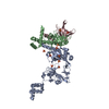
 PDBj
PDBj

 Assembly
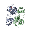


 Mass: 106.120 Da / Num. of mol.: 1 / Source method: obtained synthetically / Formula: C4H10O3
 Mass: 35.453 Da / Num. of mol.: 1 / Source method: obtained synthetically / Formula: Cl
 Mass: 18.015 Da / Num. of mol.: 290 / Source method: isolated from a natural source / Formula: H2O
 Sample preparation
 Processing