Movie
Movie Controller

[English] 日本語
 Yorodumi
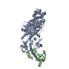
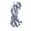
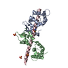
- PDB-7yv9: Cryo-EM structure of full-length Myosin Va in the autoinhibited state -
+ Open data

- Basic information
| Entry | Database: PDB / ID: 7yv9 | ||||||
|---|---|---|---|---|---|---|---|
| Title | Cryo-EM structure of full-length Myosin Va in the autoinhibited state | ||||||
 Components Components |
| ||||||
 Keywords Keywords | MOTOR PROTEIN / intracellular trafficking / molecular motor / myosin / autoinhibition | ||||||
| Function / homology |  Function and homology information Function and homology informationphenol-containing compound metabolic process / CaMK IV-mediated phosphorylation of CREB / Cam-PDE 1 activation / CREB1 phosphorylation through the activation of CaMKII/CaMKK/CaMKIV cascasde / Glycogen breakdown (glycogenolysis) / Activation of RAC1 downstream of NMDARs / Sodium/Calcium exchangers / Activation of Ca-permeable Kainate Receptor / CLEC7A (Dectin-1) induces NFAT activation / RHO GTPases activate PAKs ...phenol-containing compound metabolic process / CaMK IV-mediated phosphorylation of CREB / Cam-PDE 1 activation / CREB1 phosphorylation through the activation of CaMKII/CaMKK/CaMKIV cascasde / Glycogen breakdown (glycogenolysis) / Activation of RAC1 downstream of NMDARs / Sodium/Calcium exchangers / Activation of Ca-permeable Kainate Receptor / CLEC7A (Dectin-1) induces NFAT activation / RHO GTPases activate PAKs / Calmodulin induced events / Synthesis of IP3 and IP4 in the cytosol / Inactivation, recovery and regulation of the phototransduction cascade / Tetrahydrobiopterin (BH4) synthesis, recycling, salvage and regulation / eNOS activation / Reduction of cytosolic Ca++ levels / Calcineurin activates NFAT / Ion transport by P-type ATPases / RAF activation / VEGFR2 mediated vascular permeability / Protein methylation / vesicle transport along actin filament / RAS processing / Ca2+ pathway / FCERI mediated Ca+2 mobilization / Extra-nuclear estrogen signaling / RHO GTPases activate IQGAPs / Unblocking of NMDA receptors, glutamate binding and activation / post-Golgi vesicle-mediated transport / PKA activation / secretory granule localization / RAF/MAP kinase cascade / Smooth Muscle Contraction / filopodium tip / Platelet degranulation / High laminar flow shear stress activates signaling by PIEZO1 and PECAM1:CDH5:KDR in endothelial cells / actomyosin / Stimuli-sensing channels / : / Ion homeostasis / type 3 metabotropic glutamate receptor binding / myosin complex / microfilament motor activity / response to corticosterone / negative regulation of ryanodine-sensitive calcium-release channel activity / organelle localization by membrane tethering / : / autophagosome membrane docking / regulation of synaptic vesicle exocytosis / regulation of ryanodine-sensitive calcium-release channel activity / regulation of cardiac muscle cell action potential / presynaptic endocytosis / pigmentation / calcineurin-mediated signaling / nitric-oxide synthase binding / regulation of cell communication by electrical coupling involved in cardiac conduction / adenylate cyclase binding / protein phosphatase activator activity / regulation of synaptic vesicle endocytosis / detection of calcium ion / postsynaptic cytosol / regulation of cardiac muscle contraction / cell surface receptor signaling pathway via JAK-STAT / catalytic complex / phosphatidylinositol 3-kinase binding / calcium channel inhibitor activity / presynaptic cytosol / cellular response to interferon-beta / regulation of release of sequestered calcium ion into cytosol by sarcoplasmic reticulum / ruffle / titin binding / regulation of cardiac muscle contraction by regulation of the release of sequestered calcium ion / voltage-gated potassium channel complex / calcium channel complex / secretory granule / regulation of heart rate / calyx of Held / nitric-oxide synthase regulator activity / adenylate cyclase activator activity / protein serine/threonine kinase activator activity / regulation of cytokinesis / spindle microtubule / positive regulation of receptor signaling pathway via JAK-STAT / response to amphetamine / sarcomere / actin filament / calcium channel regulator activity / response to calcium ion / recycling endosome / cellular response to type II interferon / G2/M transition of mitotic cell cycle / Schaffer collateral - CA1 synapse / mitochondrial membrane / small GTPase binding / spindle pole / calcium-dependent protein binding / melanosome / late endosome / peroxisome / myelin sheath Similarity search - Function | ||||||
| Biological species |  | ||||||
| Method | ELECTRON MICROSCOPY / single particle reconstruction / cryo EM / Resolution: 4.78 Å | ||||||
 Authors Authors | Niu, F. / Wei, Z. | ||||||
| Funding support |  China, 1items China, 1items
| ||||||
 Citation Citation | Journal: Sci Adv / Year: 2022 Title: Autoinhibition and activation mechanisms revealed by the triangular-shaped structure of myosin Va. Authors: Fengfeng Niu / Yong Liu / Kang Sun / Shun Xu / Jiayuan Dong / Cong Yu / Kaige Yan / Zhiyi Wei / Abstract: As the prototype of unconventional myosin motor family, myosin Va (MyoVa) transport cellular cargos along actin filaments in diverse cellular processes. The off-duty MyoVa adopts a closed and ...As the prototype of unconventional myosin motor family, myosin Va (MyoVa) transport cellular cargos along actin filaments in diverse cellular processes. The off-duty MyoVa adopts a closed and autoinhibited state, which can be relieved by cargo binding. The molecular mechanisms governing the autoinhibition and activation of MyoVa remain unclear. Here, we report the cryo-electron microscopy structure of the two full-length, closed MyoVa heavy chains in complex with 12 calmodulin light chains at 4.78-Å resolution. The MyoVa adopts a triangular structure with multiple intra- and interpolypeptide chain interactions in establishing the closed state with cargo binding and adenosine triphosphatase activity inhibited. Structural, biochemical, and cellular analyses uncover an asymmetric autoinhibition mechanism, in which the cargo-binding sites in the two MyoVa heavy chains are differently protected. Thus, specific and efficient MyoVa activation requires coincident binding of multiple cargo adaptors, revealing an intricate and elegant activity regulation of the motor in response to cargos. | ||||||
| History |
|
- Structure visualization
| Structure viewer | Molecule: MolmilJmol/JSmol |
|---|
- Downloads & links
-Download
| PDBx/mmCIF format | 7yv9.cif.gz | 920.6 KB | Display | PDBx/mmCIF format |
|---|---|---|---|---|
| PDB format | pdb7yv9.ent.gz | 715.3 KB | Display | PDB format |
| PDBx/mmJSON format | 7yv9.json.gz | Tree view | PDBx/mmJSON format | |
| Others |  Other downloads Other downloads |
-Validation report
| Arichive directory | https://data.pdbj.org/pub/pdb/validation_reports/yv/7yv9ftp://data.pdbj.org/pub/pdb/validation_reports/yv/7yv9 | HTTPS FTP |
|---|
-Related structure data
| Related structure data |  34121MC M: map data used to model this data C: citing same article ( |
|---|---|
| Similar structure data |
-Links
 PDBj
PDBj


- Assembly
| Deposited unit | 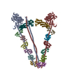
|
|---|---|
| 1 |
|
-Components
| #1: Protein | Mass: 212657.359 Da / Num. of mol.: 4 Source method: isolated from a genetically manipulated source Source: (gene. exp.)   Spodoptera frugiperda (fall armyworm) / References: UniProt: D3YZ62 Spodoptera frugiperda (fall armyworm) / References: UniProt: D3YZ62#2: Protein | Mass: 16848.605 Da / Num. of mol.: 12 / Mutation: E32Q,E68Q,E105Q,E141Q Source method: isolated from a genetically manipulated source Source: (gene. exp.) Spodoptera frugiperda (fall armyworm) / References: UniProt: P0DP26 |
|---|
-Experimental details
-Experiment
| Experiment | Method: ELECTRON MICROSCOPY |
|---|---|
| EM experiment | Aggregation state: PARTICLE / 3D reconstruction method: single particle reconstruction |
- Sample preparation
| Component | Name: Myosin Va and Calmodulin complex / Type: COMPLEX / Entity ID: all / Source: RECOMBINANT | ||||||||||||||||||||||||||||||
|---|---|---|---|---|---|---|---|---|---|---|---|---|---|---|---|---|---|---|---|---|---|---|---|---|---|---|---|---|---|---|---|
| Molecular weight | Value: 0.63 MDa / Experimental value: NO | ||||||||||||||||||||||||||||||
| Source (natural) | Organism: | ||||||||||||||||||||||||||||||
| Source (recombinant) | Organism: Spodoptera frugiperda (fall armyworm) | ||||||||||||||||||||||||||||||
| Buffer solution | pH: 7.4 | ||||||||||||||||||||||||||||||
| Buffer component |
| ||||||||||||||||||||||||||||||
| Specimen | Conc.: 0.3 mg/ml / Embedding applied: NO / Shadowing applied: NO / Staining applied: NO / Vitrification applied: YES | ||||||||||||||||||||||||||||||
| Specimen support | Grid material: COPPER / Grid type: Quantifoil R1.2/1.3 | ||||||||||||||||||||||||||||||
| Vitrification | Instrument: FEI VITROBOT MARK IV / Cryogen name: ETHANE / Humidity: 100 % / Chamber temperature: 277 K |
- Electron microscopy imaging
| Experimental equipment |  Model: Titan Krios / Image courtesy: FEI Company |
|---|---|
| Microscopy | Model: FEI TITAN KRIOS |
| Electron gun | Electron source:  FIELD EMISSION GUN / Accelerating voltage: 300 kV / Illumination mode: SPOT SCAN FIELD EMISSION GUN / Accelerating voltage: 300 kV / Illumination mode: SPOT SCAN |
| Electron lens | Mode: BRIGHT FIELD / Nominal magnification: 81000 X / Nominal defocus max: 2500 nm / Nominal defocus min: 1500 nm / Cs: 2.7 mm / Alignment procedure: BASIC |
| Specimen holder | Cryogen: NITROGEN / Specimen holder model: FEI TITAN KRIOS AUTOGRID HOLDER |
| Image recording | Average exposure time: 2 sec. / Electron dose: 50 e/Å2 / Film or detector model: GATAN K3 (6k x 4k) |
- Processing
| EM software |
| ||||||||||||||||||||||||||||||||||||||||||||
|---|---|---|---|---|---|---|---|---|---|---|---|---|---|---|---|---|---|---|---|---|---|---|---|---|---|---|---|---|---|---|---|---|---|---|---|---|---|---|---|---|---|---|---|---|---|
| CTF correction | Type: NONE | ||||||||||||||||||||||||||||||||||||||||||||
| Particle selection | Num. of particles selected: 20747000 | ||||||||||||||||||||||||||||||||||||||||||||
| Symmetry | Point symmetry: C1 (asymmetric) | ||||||||||||||||||||||||||||||||||||||||||||
| 3D reconstruction | Resolution: 4.78 Å / Resolution method: FSC 0.143 CUT-OFF / Num. of particles: 160273 / Symmetry type: POINT | ||||||||||||||||||||||||||||||||||||||||||||
| Atomic model building | B value: 167.3 / Protocol: OTHER / Space: REAL / Target criteria: Correlation coefficient | ||||||||||||||||||||||||||||||||||||||||||||
| Atomic model building |
|