Movie
Movie Controller

[English] 日本語
 Yorodumi
- PDB-7xzx: Cryo-EM structure of the nucleosome in complex with p53 DNA-bindi... -
+ Open data

- Basic information
| Entry | Database: PDB / ID: 7xzx | |||||||||||||||||||||||||||||||||
|---|---|---|---|---|---|---|---|---|---|---|---|---|---|---|---|---|---|---|---|---|---|---|---|---|---|---|---|---|---|---|---|---|---|---|
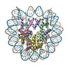
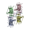
| Title | Cryo-EM structure of the nucleosome in complex with p53 DNA-binding domain | |||||||||||||||||||||||||||||||||
 Components Components |
| |||||||||||||||||||||||||||||||||
 Keywords Keywords | GENE REGURATION/DNA / Transcription factor / Tumor-suppressor / GENE REGULATION / GENE REGURATION-DNA complex | |||||||||||||||||||||||||||||||||
| Function / homology |  Function and homology information Function and homology informationnegative regulation of helicase activity / Loss of function of TP53 in cancer due to loss of tetramerization ability / Regulation of TP53 Expression / signal transduction by p53 class mediator / negative regulation of G1 to G0 transition / negative regulation of glucose catabolic process to lactate via pyruvate / Transcriptional activation of cell cycle inhibitor p21 / regulation of intrinsic apoptotic signaling pathway by p53 class mediator / negative regulation of pentose-phosphate shunt / Activation of NOXA and translocation to mitochondria ...negative regulation of helicase activity / Loss of function of TP53 in cancer due to loss of tetramerization ability / Regulation of TP53 Expression / signal transduction by p53 class mediator / negative regulation of G1 to G0 transition / negative regulation of glucose catabolic process to lactate via pyruvate / Transcriptional activation of cell cycle inhibitor p21 / regulation of intrinsic apoptotic signaling pathway by p53 class mediator / negative regulation of pentose-phosphate shunt / Activation of NOXA and translocation to mitochondria / ATP-dependent DNA/DNA annealing activity / regulation of cell cycle G2/M phase transition / oligodendrocyte apoptotic process / negative regulation of miRNA processing / intrinsic apoptotic signaling pathway in response to hypoxia / oxidative stress-induced premature senescence / regulation of tissue remodeling / positive regulation of thymocyte apoptotic process / positive regulation of mitochondrial membrane permeability / germ cell nucleus / regulation of fibroblast apoptotic process / bone marrow development / cellular response to actinomycin D / circadian behavior / regulation of mitochondrial membrane permeability involved in apoptotic process / histone deacetylase regulator activity / positive regulation of programmed necrotic cell death / : / RUNX3 regulates CDKN1A transcription / T cell proliferation involved in immune response / TP53 Regulates Transcription of Death Receptors and Ligands / Activation of PUMA and translocation to mitochondria / TP53 regulates transcription of additional cell cycle genes whose exact role in the p53 pathway remain uncertain / mRNA transcription / negative regulation of glial cell proliferation / negative regulation of neuroblast proliferation / regulation of DNA damage response, signal transduction by p53 class mediator / Regulation of TP53 Activity through Association with Co-factors / Formation of Senescence-Associated Heterochromatin Foci (SAHF) / mitochondrial DNA repair / T cell lineage commitment / thymocyte apoptotic process / ER overload response / TP53 Regulates Transcription of Caspase Activators and Caspases / cardiac septum morphogenesis / necroptotic process / B cell lineage commitment / entrainment of circadian clock by photoperiod / negative regulation of DNA replication / Zygotic genome activation (ZGA) / negative regulation of mitophagy / TP53 Regulates Transcription of Genes Involved in Cytochrome C Release / PI5P Regulates TP53 Acetylation / positive regulation of release of cytochrome c from mitochondria / neuroblast proliferation / Association of TriC/CCT with target proteins during biosynthesis / negative regulation of telomere maintenance via telomerase / SUMOylation of transcription factors / TP53 regulates transcription of several additional cell death genes whose specific roles in p53-dependent apoptosis remain uncertain / rRNA transcription / negative regulation of reactive oxygen species metabolic process / intrinsic apoptotic signaling pathway by p53 class mediator / TFIID-class transcription factor complex binding / Transcriptional Regulation by VENTX / cellular response to UV-C / replicative senescence / viral process / intrinsic apoptotic signaling pathway in response to endoplasmic reticulum stress / hematopoietic stem cell differentiation / embryonic organ development / intrinsic apoptotic signaling pathway in response to DNA damage by p53 class mediator / Pyroptosis / positive regulation of RNA polymerase II transcription preinitiation complex assembly / chromosome organization / general transcription initiation factor binding / positive regulation of execution phase of apoptosis / type II interferon-mediated signaling pathway / hematopoietic progenitor cell differentiation / negative regulation of stem cell proliferation / TP53 Regulates Transcription of Genes Involved in G1 Cell Cycle Arrest / response to X-ray / negative regulation of tumor necrosis factor-mediated signaling pathway / somitogenesis / negative regulation of fibroblast proliferation / core promoter sequence-specific DNA binding / glial cell proliferation / cis-regulatory region sequence-specific DNA binding / cellular response to glucose starvation / mitophagy / Regulation of TP53 Activity through Acetylation / negative regulation of megakaryocyte differentiation / protein localization to CENP-A containing chromatin / Chromatin modifying enzymes / negative regulation of proteolysis / mitotic G1 DNA damage checkpoint signaling / positive regulation of intrinsic apoptotic signaling pathway / Replacement of protamines by nucleosomes in the male pronucleus / response to salt stress / CENP-A containing nucleosome / cardiac muscle cell apoptotic process Similarity search - Function | |||||||||||||||||||||||||||||||||
| Biological species |  Homo sapiens (human) Homo sapiens (human)synthetic construct (others) | |||||||||||||||||||||||||||||||||
| Method | ELECTRON MICROSCOPY / single particle reconstruction / cryo EM / Resolution: 4.53 Å | |||||||||||||||||||||||||||||||||
 Authors Authors | Nishimura, M. / Nozawa, K. / Takizawa, Y. / Kurumizaka, H. | |||||||||||||||||||||||||||||||||
| Funding support |  Japan, 10items Japan, 10items
| |||||||||||||||||||||||||||||||||
 Citation Citation | Journal: PNAS Nexus / Year: 2022 Title: Structural basis for p53 binding to its nucleosomal target DNA sequence. Authors: Masahiro Nishimura / Yoshimasa Takizawa / Kayo Nozawa / Hitoshi Kurumizaka / Abstract: The tumor suppressor p53 functions as a pioneer transcription factor that binds a nucleosomal target DNA sequence. However, the mechanism by which p53 binds to its target DNA in the nucleosome ...The tumor suppressor p53 functions as a pioneer transcription factor that binds a nucleosomal target DNA sequence. However, the mechanism by which p53 binds to its target DNA in the nucleosome remains elusive. Here we report the cryo-electron microscopy structures of the p53 DNA-binding domain and the full-length p53 protein complexed with a nucleosome containing the 20 base-pair target DNA sequence of p53 (p53BS). In the p53-nucleosome structures, the p53 DNA-binding domain forms a tetramer and specifically binds to the p53BS DNA, located near the entry/exit region of the nucleosome. The nucleosomal position of the p53BS DNA is within the genomic p21 promoter region. The p53 binding peels the DNA from the histone surface, and drastically changes the DNA path around the p53BS on the nucleosome. The C-terminal domain of p53 also binds to the DNA around the center and linker DNA regions of the nucleosome, as revealed by hydroxyl radical footprinting. These results provide important structural information for understanding the mechanism by which p53 binds the nucleosome and changes the chromatin structure for gene activation. | |||||||||||||||||||||||||||||||||
| History |
|
- Structure visualization
| Structure viewer | Molecule: MolmilJmol/JSmol |
|---|
- Downloads & links
-Download
| PDBx/mmCIF format | 7xzx.cif.gz | 462 KB | Display | PDBx/mmCIF format |
|---|---|---|---|---|
| PDB format | pdb7xzx.ent.gz | 359 KB | Display | PDB format |
| PDBx/mmJSON format | 7xzx.json.gz | Tree view | PDBx/mmJSON format | |
| Others |  Other downloads Other downloads |
-Validation report
| Arichive directory | https://data.pdbj.org/pub/pdb/validation_reports/xz/7xzxftp://data.pdbj.org/pub/pdb/validation_reports/xz/7xzx | HTTPS FTP |
|---|
-Related structure data
| Related structure data |  33533MC 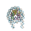 7xzyC 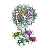 7xzzC 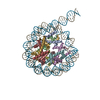 7y00C M: map data used to model this data C: citing same article ( |
|---|---|
| Similar structure data |


-Links
 PDBj
PDBj


- Assembly
| Deposited unit | 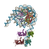
|
|---|---|
| 1 |
|
-Components
-Protein , 5 types, 12 molecules AEBFCGDHKLMN
| #1: Protein | Mass: 15719.445 Da / Num. of mol.: 2 Source method: isolated from a genetically manipulated source Source: (gene. exp.) Homo sapiens (human) / Gene: H3C1 / Production host:  #2: Protein | Mass: 11676.703 Da / Num. of mol.: 2 Source method: isolated from a genetically manipulated source Source: (gene. exp.) Homo sapiens (human) / Gene: H4C1 / Production host: #3: Protein | Mass: 14447.825 Da / Num. of mol.: 2 Source method: isolated from a genetically manipulated source Source: (gene. exp.) Homo sapiens (human) / Gene: HIST1H2AE / Production host: #4: Protein | Mass: 14217.516 Da / Num. of mol.: 2 Source method: isolated from a genetically manipulated source Source: (gene. exp.) Homo sapiens (human) / Gene: HIST1H2BJ / Production host: #7: Protein | Mass: 22659.750 Da / Num. of mol.: 4 Source method: isolated from a genetically manipulated source Source: (gene. exp.) Homo sapiens (human) / Gene: TP53, P53 / Production host: |
|---|
-DNA chain , 2 types, 2 molecules IJ
| #5: DNA chain | Mass: 59417.824 Da / Num. of mol.: 1 / Source method: obtained synthetically / Source: (synth.) synthetic construct (others) |
|---|---|
| #6: DNA chain | Mass: 59760.027 Da / Num. of mol.: 1 / Source method: obtained synthetically / Source: (synth.) synthetic construct (others) |
-Experimental details
-Experiment
| Experiment | Method: ELECTRON MICROSCOPY |
|---|---|
| EM experiment | Aggregation state: PARTICLE / 3D reconstruction method: single particle reconstruction |
- Sample preparation
| Component |
| ||||||||||||||||||||||||
|---|---|---|---|---|---|---|---|---|---|---|---|---|---|---|---|---|---|---|---|---|---|---|---|---|---|
| Molecular weight | Value: 0.329 MDa / Experimental value: NO | ||||||||||||||||||||||||
| Source (natural) | Organism: Homo sapiens (human) | ||||||||||||||||||||||||
| Source (recombinant) | Organism: | ||||||||||||||||||||||||
| Buffer solution | pH: 8 | ||||||||||||||||||||||||
| Buffer component |
| ||||||||||||||||||||||||
| Specimen | Conc.: 0.5 mg/ml / Embedding applied: NO / Shadowing applied: NO / Staining applied: NO / Vitrification applied: YES | ||||||||||||||||||||||||
| Specimen support | Grid material: COPPER / Grid mesh size: 200 divisions/in. / Grid type: Quantifoil R1.2/1.3 | ||||||||||||||||||||||||
| Vitrification | Instrument: FEI VITROBOT MARK IV / Cryogen name: ETHANE / Humidity: 100 % / Chamber temperature: 289.15 K |
- Electron microscopy imaging
| Experimental equipment |  Model: Titan Krios / Image courtesy: FEI Company |
|---|---|
| Microscopy | Model: FEI TITAN KRIOS |
| Electron gun | Electron source:  FIELD EMISSION GUN / Accelerating voltage: 300 kV / Illumination mode: FLOOD BEAM FIELD EMISSION GUN / Accelerating voltage: 300 kV / Illumination mode: FLOOD BEAM |
| Electron lens | Mode: BRIGHT FIELD / Nominal defocus max: 2500 nm / Nominal defocus min: 1000 nm |
| Image recording | Electron dose: 60 e/Å2 / Film or detector model: GATAN K3 BIOQUANTUM (6k x 4k) |
- Processing
| EM software |
| ||||||||||||||||||||||||||||||||||||
|---|---|---|---|---|---|---|---|---|---|---|---|---|---|---|---|---|---|---|---|---|---|---|---|---|---|---|---|---|---|---|---|---|---|---|---|---|---|
| CTF correction | Type: PHASE FLIPPING AND AMPLITUDE CORRECTION | ||||||||||||||||||||||||||||||||||||
| Particle selection | Num. of particles selected: 2452876 | ||||||||||||||||||||||||||||||||||||
| Symmetry | Point symmetry: C1 (asymmetric) | ||||||||||||||||||||||||||||||||||||
| 3D reconstruction | Resolution: 4.53 Å / Resolution method: FSC 0.143 CUT-OFF / Num. of particles: 53958 / Symmetry type: POINT | ||||||||||||||||||||||||||||||||||||
| Atomic model building | Space: REAL | ||||||||||||||||||||||||||||||||||||
| Atomic model building |
|