Movie
Movie Controller

[English] 日本語
 Yorodumi
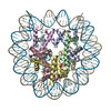
- PDB-7xzy: Cryo-EM structure of the nucleosome containing 193 base-pair DNA ... -
+ Open data

- Basic information
| Entry | Database: PDB / ID: 7xzy | |||||||||||||||||||||||||||||||||
|---|---|---|---|---|---|---|---|---|---|---|---|---|---|---|---|---|---|---|---|---|---|---|---|---|---|---|---|---|---|---|---|---|---|---|
| Title | Cryo-EM structure of the nucleosome containing 193 base-pair DNA with a p53 target sequence | |||||||||||||||||||||||||||||||||
 Components Components |
| |||||||||||||||||||||||||||||||||
 Keywords Keywords | GENE REGURATION/DNA / Transcription factor / Tumor-suppressor / GENE REGURATION-DNA complex | |||||||||||||||||||||||||||||||||
| Function / homology |  Function and homology information Function and homology informationnegative regulation of tumor necrosis factor-mediated signaling pathway / negative regulation of megakaryocyte differentiation / protein localization to CENP-A containing chromatin / Chromatin modifying enzymes / Replacement of protamines by nucleosomes in the male pronucleus / CENP-A containing nucleosome / Packaging Of Telomere Ends / Recognition and association of DNA glycosylase with site containing an affected purine / Cleavage of the damaged purine / epigenetic regulation of gene expression ...negative regulation of tumor necrosis factor-mediated signaling pathway / negative regulation of megakaryocyte differentiation / protein localization to CENP-A containing chromatin / Chromatin modifying enzymes / Replacement of protamines by nucleosomes in the male pronucleus / CENP-A containing nucleosome / Packaging Of Telomere Ends / Recognition and association of DNA glycosylase with site containing an affected purine / Cleavage of the damaged purine / epigenetic regulation of gene expression / Deposition of new CENPA-containing nucleosomes at the centromere / telomere organization / Interleukin-7 signaling / Recognition and association of DNA glycosylase with site containing an affected pyrimidine / Cleavage of the damaged pyrimidine / RNA Polymerase I Promoter Opening / Inhibition of DNA recombination at telomere / Assembly of the ORC complex at the origin of replication / Meiotic synapsis / SUMOylation of chromatin organization proteins / Regulation of endogenous retroelements by the Human Silencing Hub (HUSH) complex / DNA methylation / Condensation of Prophase Chromosomes / Chromatin modifications during the maternal to zygotic transition (MZT) / SIRT1 negatively regulates rRNA expression / HCMV Late Events / ERCC6 (CSB) and EHMT2 (G9a) positively regulate rRNA expression / PRC2 methylates histones and DNA / Regulation of endogenous retroelements by KRAB-ZFP proteins / Defective pyroptosis / innate immune response in mucosa / HDACs deacetylate histones / Regulation of endogenous retroelements by Piwi-interacting RNAs (piRNAs) / RNA Polymerase I Promoter Escape / Nonhomologous End-Joining (NHEJ) / lipopolysaccharide binding / Transcriptional regulation by small RNAs / HDMs demethylate histones / Formation of the beta-catenin:TCF transactivating complex / Activated PKN1 stimulates transcription of AR (androgen receptor) regulated genes KLK2 and KLK3 / RUNX1 regulates genes involved in megakaryocyte differentiation and platelet function / NoRC negatively regulates rRNA expression / Negative Regulation of CDH1 Gene Transcription / G2/M DNA damage checkpoint / PKMTs methylate histone lysines / B-WICH complex positively regulates rRNA expression / DNA Damage/Telomere Stress Induced Senescence / Meiotic recombination / Pre-NOTCH Transcription and Translation / Activation of anterior HOX genes in hindbrain development during early embryogenesis / Transcriptional regulation of granulopoiesis / RMTs methylate histone arginines / Metalloprotease DUBs / HCMV Early Events / structural constituent of chromatin / UCH proteinases / nucleosome / antimicrobial humoral immune response mediated by antimicrobial peptide / nucleosome assembly / HATs acetylate histones / E3 ubiquitin ligases ubiquitinate target proteins / antibacterial humoral response / heterochromatin formation / Recruitment and ATM-mediated phosphorylation of repair and signaling proteins at DNA double strand breaks / Factors involved in megakaryocyte development and platelet production / MLL4 and MLL3 complexes regulate expression of PPARG target genes in adipogenesis and hepatic steatosis / chromatin organization / RUNX1 regulates transcription of genes involved in differentiation of HSCs / Processing of DNA double-strand break ends / Senescence-Associated Secretory Phenotype (SASP) / Oxidative Stress Induced Senescence / Estrogen-dependent gene expression / killing of cells of another organism / defense response to Gram-negative bacterium / chromosome, telomeric region / Ub-specific processing proteases / defense response to Gram-positive bacterium / cadherin binding / Amyloid fiber formation / protein heterodimerization activity / negative regulation of cell population proliferation / protein-containing complex / : / DNA binding / RNA binding / extracellular exosome / extracellular region / nucleoplasm / membrane / nucleus / cytosol Similarity search - Function | |||||||||||||||||||||||||||||||||
| Biological species |  Homo sapiens (human) Homo sapiens (human)synthetic construct (others) | |||||||||||||||||||||||||||||||||
| Method | ELECTRON MICROSCOPY / single particle reconstruction / cryo EM / Resolution: 3.97 Å | |||||||||||||||||||||||||||||||||
 Authors Authors | Nishimura, M. / Nozawa, K. / Takizawa, Y. / Kurumizaka, H. | |||||||||||||||||||||||||||||||||
| Funding support |  Japan, 10items Japan, 10items
| |||||||||||||||||||||||||||||||||
 Citation Citation | Journal: PNAS Nexus / Year: 2022 Title: Structural basis for p53 binding to its nucleosomal target DNA sequence. Authors: Masahiro Nishimura / Yoshimasa Takizawa / Kayo Nozawa / Hitoshi Kurumizaka / Abstract: The tumor suppressor p53 functions as a pioneer transcription factor that binds a nucleosomal target DNA sequence. However, the mechanism by which p53 binds to its target DNA in the nucleosome ...The tumor suppressor p53 functions as a pioneer transcription factor that binds a nucleosomal target DNA sequence. However, the mechanism by which p53 binds to its target DNA in the nucleosome remains elusive. Here we report the cryo-electron microscopy structures of the p53 DNA-binding domain and the full-length p53 protein complexed with a nucleosome containing the 20 base-pair target DNA sequence of p53 (p53BS). In the p53-nucleosome structures, the p53 DNA-binding domain forms a tetramer and specifically binds to the p53BS DNA, located near the entry/exit region of the nucleosome. The nucleosomal position of the p53BS DNA is within the genomic p21 promoter region. The p53 binding peels the DNA from the histone surface, and drastically changes the DNA path around the p53BS on the nucleosome. The C-terminal domain of p53 also binds to the DNA around the center and linker DNA regions of the nucleosome, as revealed by hydroxyl radical footprinting. These results provide important structural information for understanding the mechanism by which p53 binds the nucleosome and changes the chromatin structure for gene activation. | |||||||||||||||||||||||||||||||||
| History |
|
- Structure visualization
| Structure viewer | Molecule: MolmilJmol/JSmol |
|---|
- Downloads & links
-Download
| PDBx/mmCIF format | 7xzy.cif.gz | 305.1 KB | Display | PDBx/mmCIF format |
|---|---|---|---|---|
| PDB format | pdb7xzy.ent.gz | 227.3 KB | Display | PDB format |
| PDBx/mmJSON format | 7xzy.json.gz | Tree view | PDBx/mmJSON format | |
| Others |  Other downloads Other downloads |
-Validation report
| Arichive directory | https://data.pdbj.org/pub/pdb/validation_reports/xz/7xzyftp://data.pdbj.org/pub/pdb/validation_reports/xz/7xzy | HTTPS FTP |
|---|
-Related structure data
| Related structure data |  33534MC 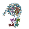 7xzxC 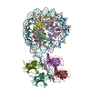 7xzzC 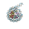 7y00C M: map data used to model this data C: citing same article ( |
|---|---|
| Similar structure data |


-Links
 PDBj
PDBj


- Assembly
| Deposited unit | 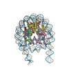
|
|---|---|
| 1 |
|
-Components
-Protein , 4 types, 8 molecules AEBFCGDH
| #1: Protein | Mass: 15719.445 Da / Num. of mol.: 2 Source method: isolated from a genetically manipulated source Source: (gene. exp.) Homo sapiens (human) / Gene: H3C1 / Production host:  #2: Protein | Mass: 11676.703 Da / Num. of mol.: 2 Source method: isolated from a genetically manipulated source Source: (gene. exp.) Homo sapiens (human) / Gene: H4C1 / Production host: #3: Protein | Mass: 14447.825 Da / Num. of mol.: 2 Source method: isolated from a genetically manipulated source Source: (gene. exp.) Homo sapiens (human) / Gene: H2AC4 / Production host: #4: Protein | Mass: 14217.516 Da / Num. of mol.: 2 Source method: isolated from a genetically manipulated source Source: (gene. exp.) Homo sapiens (human) / Gene: H2BC11 / Production host: |
|---|
-DNA chain , 2 types, 2 molecules IJ
| #5: DNA chain | Mass: 59417.824 Da / Num. of mol.: 1 / Source method: obtained synthetically / Source: (synth.) synthetic construct (others) |
|---|---|
| #6: DNA chain | Mass: 59760.027 Da / Num. of mol.: 1 / Source method: obtained synthetically / Source: (synth.) synthetic construct (others) |
-Experimental details
-Experiment
| Experiment | Method: ELECTRON MICROSCOPY |
|---|---|
| EM experiment | Aggregation state: PARTICLE / 3D reconstruction method: single particle reconstruction |
- Sample preparation
| Component |
| ||||||||||||||||||||||||
|---|---|---|---|---|---|---|---|---|---|---|---|---|---|---|---|---|---|---|---|---|---|---|---|---|---|
| Molecular weight | Value: 0.239 MDa / Experimental value: NO | ||||||||||||||||||||||||
| Source (natural) | Organism: Homo sapiens (human) | ||||||||||||||||||||||||
| Source (recombinant) | Organism: | ||||||||||||||||||||||||
| Buffer solution | pH: 8 | ||||||||||||||||||||||||
| Buffer component |
| ||||||||||||||||||||||||
| Specimen | Conc.: 0.5 mg/ml / Embedding applied: NO / Shadowing applied: NO / Staining applied: NO / Vitrification applied: YES | ||||||||||||||||||||||||
| Vitrification | Instrument: FEI VITROBOT MARK IV / Cryogen name: ETHANE / Humidity: 100 % / Chamber temperature: 289.15 K |
- Electron microscopy imaging
| Experimental equipment |  Model: Titan Krios / Image courtesy: FEI Company |
|---|---|
| Microscopy | Model: FEI TITAN KRIOS |
| Electron gun | Electron source:  FIELD EMISSION GUN / Accelerating voltage: 300 kV / Illumination mode: FLOOD BEAM FIELD EMISSION GUN / Accelerating voltage: 300 kV / Illumination mode: FLOOD BEAM |
| Electron lens | Mode: BRIGHT FIELD / Nominal defocus max: 2500 nm / Nominal defocus min: 1000 nm |
| Image recording | Electron dose: 60 e/Å2 / Film or detector model: GATAN K3 BIOQUANTUM (6k x 4k) |
- Processing
| EM software |
| ||||||||||||||||||||||||||||||||
|---|---|---|---|---|---|---|---|---|---|---|---|---|---|---|---|---|---|---|---|---|---|---|---|---|---|---|---|---|---|---|---|---|---|
| CTF correction | Type: PHASE FLIPPING AND AMPLITUDE CORRECTION | ||||||||||||||||||||||||||||||||
| Particle selection | Num. of particles selected: 2452876 | ||||||||||||||||||||||||||||||||
| 3D reconstruction | Resolution: 3.97 Å / Resolution method: FSC 0.143 CUT-OFF / Num. of particles: 161202 / Symmetry type: POINT | ||||||||||||||||||||||||||||||||
| Atomic model building | Space: REAL | ||||||||||||||||||||||||||||||||
| Atomic model building | PDB-ID: 7OHC Accession code: 7OHC / Source name: PDB / Type: experimental model |