 Movie
Movie Controller

[English] 日本語
 Yorodumi
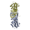
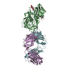
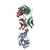
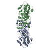
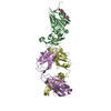
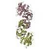
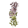
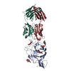
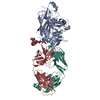
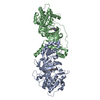
- PDB-7wue: Crystal structure of SARS-CoV-2 Receptor Binding Domain in comple... -
+ Open data

- Basic information
| Entry | Database: PDB / ID: 7wue | ||||||
|---|---|---|---|---|---|---|---|
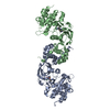
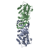
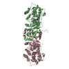
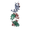
| Title | Crystal structure of SARS-CoV-2 Receptor Binding Domain in complex with the monoclonal antibody m31A7 | ||||||
 Components Components |
| ||||||
 Keywords Keywords | VIRAL PROTEIN/IMMUNE SYSTEM / SARS-CoV-2 / Spike / receptor binding domain / m31A7 / monoclonal antibody / VIRAL PROTEIN / VIRAL PROTEIN-IMMUNE SYSTEM complex | ||||||
| Function / homology |  Function and homology information Function and homology informationMaturation of spike protein / Translation of Structural Proteins / Virion Assembly and Release / host cell surface / host extracellular space / viral translation / symbiont-mediated-mediated suppression of host tetherin activity / Induction of Cell-Cell Fusion / structural constituent of virion / membrane fusion ...Maturation of spike protein / Translation of Structural Proteins / Virion Assembly and Release / host cell surface / host extracellular space / viral translation / symbiont-mediated-mediated suppression of host tetherin activity / Induction of Cell-Cell Fusion / structural constituent of virion / membrane fusion / entry receptor-mediated virion attachment to host cell / Attachment and Entry / host cell endoplasmic reticulum-Golgi intermediate compartment membrane / positive regulation of viral entry into host cell / receptor-mediated virion attachment to host cell / host cell surface receptor binding / symbiont-mediated suppression of host innate immune response / receptor ligand activity / endocytosis involved in viral entry into host cell / fusion of virus membrane with host plasma membrane / fusion of virus membrane with host endosome membrane / viral envelope / virion attachment to host cell / SARS-CoV-2 activates/modulates innate and adaptive immune responses / host cell plasma membrane / virion membrane / identical protein binding / membrane / plasma membrane Similarity search - Function | ||||||
| Biological species |   Severe acute respiratory syndrome coronavirus 2 Severe acute respiratory syndrome coronavirus 2 | ||||||
| Method |  X-RAY DIFFRACTION / SYNCHROTRON / MOLECULAR REPLACEMENT / Resolution: 3.2 Å X-RAY DIFFRACTION / SYNCHROTRON / MOLECULAR REPLACEMENT / Resolution: 3.2 Å | ||||||
 Authors Authors | Mohapatra, A. | ||||||
| Funding support |  Taiwan, 1items Taiwan, 1items
| ||||||
 Citation Citation | Journal: Sci Transl Med / Year: 2022 Title: Vaccination with SARS-CoV-2 spike protein lacking glycan shields elicits enhanced protective responses in animal models. Authors: Han-Yi Huang / Hsin-Yu Liao / Xiaorui Chen / Szu-Wen Wang / Cheng-Wei Cheng / Md Shahed-Al-Mahmud / Yo-Min Liu / Arpita Mohapatra / Ting-Hua Chen / Jennifer M Lo / Yi-Min Wu / Hsiu-Hua Ma / ...Authors: Han-Yi Huang / Hsin-Yu Liao / Xiaorui Chen / Szu-Wen Wang / Cheng-Wei Cheng / Md Shahed-Al-Mahmud / Yo-Min Liu / Arpita Mohapatra / Ting-Hua Chen / Jennifer M Lo / Yi-Min Wu / Hsiu-Hua Ma / Yi-Hsuan Chang / Ho-Yang Tsai / Yu-Chi Chou / Yi-Ping Hsueh / Ching-Yen Tsai / Pau-Yi Huang / Sui-Yuan Chang / Tai-Ling Chao / Han-Chieh Kao / Ya-Min Tsai / Yen-Hui Chen / Chung-Yi Wu / Jia-Tsrong Jan / Ting-Jen Rachel Cheng / Kuo-I Lin / Che Ma / Chi-Huey Wong /  Abstract: A major challenge to end the pandemic caused by severe acute respiratory syndrome coronavirus 2 (SARS-CoV-2) is to develop a broadly protective vaccine that elicits long-term immunity. As the key ...A major challenge to end the pandemic caused by severe acute respiratory syndrome coronavirus 2 (SARS-CoV-2) is to develop a broadly protective vaccine that elicits long-term immunity. As the key immunogen, the viral surface spike (S) protein is frequently mutated, and conserved epitopes are shielded by glycans. Here, we revealed that S protein glycosylation has site-differential effects on viral infectivity. We found that S protein generated by lung epithelial cells has glycoforms associated with increased infectivity. Compared to the fully glycosylated S protein, immunization of S protein with N-glycans trimmed to the mono-GlcNAc-decorated state (S) elicited stronger immune responses and better protection for human angiotensin-converting enzyme 2 (hACE2) transgenic mice against variants of concern (VOCs). In addition, a broadly neutralizing monoclonal antibody was identified from S-immunized mice that could neutralize wild-type SARS-CoV-2 and VOCs with subpicomolar potency. Together, these results demonstrate that removal of glycan shields to better expose the conserved sequences has the potential to be an effective and simple approach for developing a broadly protective SARS-CoV-2 vaccine. | ||||||
| History |
|
- Structure visualization
| Structure viewer | Molecule: MolmilJmol/JSmol |
|---|
- Downloads & links
-Download
| PDBx/mmCIF format | 7wue.cif.gz | 261.1 KB | Display | PDBx/mmCIF format |
|---|---|---|---|---|
| PDB format | pdb7wue.ent.gz | 207.3 KB | Display | PDB format |
| PDBx/mmJSON format | 7wue.json.gz | Tree view | PDBx/mmJSON format | |
| Others |  Other downloads Other downloads |
-Validation report
| Summary document | 7wue_validation.pdf.gz | 1.6 MB | Display | wwPDB validaton report |
|---|---|---|---|---|
| Full document | 7wue_full_validation.pdf.gz | 1.6 MB | Display | |
| Data in XML | 7wue_validation.xml.gz | 48 KB | Display | |
| Data in CIF | 7wue_validation.cif.gz | 65.6 KB | Display | |
| Arichive directory | https://data.pdbj.org/pub/pdb/validation_reports/wu/7wueftp://data.pdbj.org/pub/pdb/validation_reports/wu/7wue | HTTPS FTP |
-Related structure data
| Related structure data |  7wuhC  7c01S S: Starting model for refinement C: citing same article ( |
|---|---|
| Similar structure data |


-Links
 PDBj
PDBj


- Assembly
| Deposited unit | 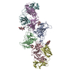
| ||||||||
|---|---|---|---|---|---|---|---|---|---|
| 1 | 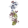
| ||||||||
| 2 | 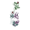
| ||||||||
| Unit cell |
|
-Components
| #1: Protein | Mass: 21873.496 Da / Num. of mol.: 2 Source method: isolated from a genetically manipulated source Source: (gene. exp.) Severe acute respiratory syndrome coronavirus 2Gene: S, 2 / Production host:  Homo sapiens (human) / References: UniProt: P0DTC2 Homo sapiens (human) / References: UniProt: P0DTC2#2: Antibody | Mass: 25621.902 Da / Num. of mol.: 2 Source method: isolated from a genetically manipulated source Source: (gene. exp.) Homo sapiens (human)#3: Antibody | Mass: 26564.006 Da / Num. of mol.: 2 Source method: isolated from a genetically manipulated source Source: (gene. exp.) Homo sapiens (human)#4: Polysaccharide | Source method: isolated from a genetically manipulated source #5: Polysaccharide | Source method: isolated from a genetically manipulated source Has ligand of interest | Y | Has protein modification | Y | |
|---|
-Experimental details
-Experiment
| Experiment | Method: X-RAY DIFFRACTION / Number of used crystals: 1 |
|---|
- Sample preparation
| Crystal | Density Matthews: 3.35 Å3/Da / Density % sol: 63.26 % |
|---|---|
| Crystal grow | Temperature: 293.15 K / Method: vapor diffusion, hanging drop / pH: 4.6 / Details: 2.0 M Ammonia sulfate, 0.1M sodium acetate pH 4.6 |
-Data collection
| Diffraction | Mean temperature: 100 K / Serial crystal experiment: N | ||||||||||||||||||||||||||||||
|---|---|---|---|---|---|---|---|---|---|---|---|---|---|---|---|---|---|---|---|---|---|---|---|---|---|---|---|---|---|---|---|
| Diffraction source | Source: SYNCHROTRON / Site: NSRRC / Beamline: BL15A1 / Wavelength: 1 Å | ||||||||||||||||||||||||||||||
| Detector | Type: RAYONIX MX300HE / Detector: CCD / Date: Dec 8, 2021 | ||||||||||||||||||||||||||||||
| Radiation | Monochromator: LN2-Cooled, Fixed-Exit Double Crystal Monochromator Protocol: SINGLE WAVELENGTH / Monochromatic (M) / Laue (L): M / Scattering type: x-ray | ||||||||||||||||||||||||||||||
| Radiation wavelength | Wavelength: 1 Å / Relative weight: 1 | ||||||||||||||||||||||||||||||
| Reflection | Resolution: 3.2→48.91 Å / Num. obs: 33348 / % possible obs: 99.3 % / Redundancy: 3.4 % / Biso Wilson estimate: 66.59 Å2 / CC1/2: 0.983 / Rmerge(I) obs: 0.182 / Rpim(I) all: 0.11 / Rrim(I) all: 0.214 / Net I/σ(I): 5.7 / Num. measured all: 114117 / Scaling rejects: 125 | ||||||||||||||||||||||||||||||
| Reflection shell | Diffraction-ID: 1
|
- Processing
| Software |
| |||||||||||||||||||||||||||||||||||||||||||||||||||||||||||||||||||||||||||||||||||||||||||
|---|---|---|---|---|---|---|---|---|---|---|---|---|---|---|---|---|---|---|---|---|---|---|---|---|---|---|---|---|---|---|---|---|---|---|---|---|---|---|---|---|---|---|---|---|---|---|---|---|---|---|---|---|---|---|---|---|---|---|---|---|---|---|---|---|---|---|---|---|---|---|---|---|---|---|---|---|---|---|---|---|---|---|---|---|---|---|---|---|---|---|---|---|
| Refinement | Method to determine structure: MOLECULAR REPLACEMENT Starting model: 7C01 Resolution: 3.2→47.61 Å / SU ML: 0.52 / Cross valid method: THROUGHOUT / σ(F): 1.36 / Phase error: 30.62 / Stereochemistry target values: ML
| |||||||||||||||||||||||||||||||||||||||||||||||||||||||||||||||||||||||||||||||||||||||||||
| Solvent computation | Shrinkage radii: 0.9 Å / VDW probe radii: 1.11 Å / Solvent model: FLAT BULK SOLVENT MODEL | |||||||||||||||||||||||||||||||||||||||||||||||||||||||||||||||||||||||||||||||||||||||||||
| Displacement parameters | Biso max: 142.24 Å2 / Biso mean: 64.7867 Å2 / Biso min: 25.02 Å2 | |||||||||||||||||||||||||||||||||||||||||||||||||||||||||||||||||||||||||||||||||||||||||||
| Refinement step | Cycle: final / Resolution: 3.2→47.61 Å
| |||||||||||||||||||||||||||||||||||||||||||||||||||||||||||||||||||||||||||||||||||||||||||
| LS refinement shell | Refine-ID: X-RAY DIFFRACTION / Rfactor Rfree error: 0 / Total num. of bins used: 12
|
